Introduction
Upper extremity and neck pain (NSUEP) is the constellation of disorders caused by neck, shoulder or lower extremity pain. NSUEP is defined as having at least one area of pain in the neck upper extremities, or shoulders. The self-reported surveys of patients show that the frequency of upper and neck pain in the first month was 44 percent. 1 Previous studies have also revealed that the 12 month-period incidence of neck pain is 31.4 percentage the shoulder pain rate is 30.3 percent and wrist pain is 11.2 percent and hand or wrist pain is 17.5 percentage in the overall populace. 2
|
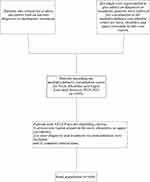
Image 1 Diagram of the flow of the patient’s selection. |
|
2. Age distribution of 444 patients at the time of their visit. The patients who develop NSUEP typically are between 51 and 60 and older (157 patient, 35.4%). |
|
FIGURE 3 The pain regions distribution for patients suffering from NSUEP. The most commonly affected region for tenderness was on the upper extremities. |
|
Figure 4. The distribution of symptoms in patients suffering from NSUEP. The weakness, numbness, and inactivity are all common symptoms, in addition to discomfort. |
|
Figure 5. Sign distribution among patients suffering from NSUEP. Hypoesthesia, Hoffmann’s sign muscles weakness, muscle atrophy and Spurling’s test are typical symptoms. |
|
FIGURE 6 A single patient are classified by department. |
|
Figure 7. Cervical spondylosis has been classified into five kinds by the diagnostic category. |
|
Table 1. Indicative Frequency as well as Percentage of clinical signs of NSUEP in the Regions of Pain |
|
Table 2. Diagnosis Rates for NSUEP based on the clinical Signs |
|
Table 3 Patients with Cervical Radiculopathy and Compound Diagnosis |
Machino et al 3 found that shoulder and neck discomfort was associated with the poor health-related quality of life for a middle-aged, community-based population. Patients suffering from NSUEP were affected by various symptoms, which resulted in a lower quality of life as well as significant social costs.4 In Sweden it is estimated that shoulder and neck issues make up 18 percent from all disabilities payments.5 Additionally to that, the frequent occurrence of these problems and the complexity of their causes leads to numerous patients who have no diagnosis.6 Treatments included non-pharmacological as well as pharmacological therapies7 and invasive surgical interventions in cases where an important pathology is involved.8
The study examined the prospective clinical records of patients suffering from NSUEP who attended the multidisciplinary consult center for shoulder, neck and upper extremity pain from 2014 between 2014 and 2021. The aim was to provide detailed clinical characteristics and diagnoses for NSUEP in a single clinic and to increase understanding among clinicians of this disorder.
Materials and Methods
All patients received treatment in the multidisciplinary clinic for shoulder, neck, and upper extremity pains at China-Japan Union Hospital at Jilin University between April 2014 until July 2021. The clinical data was retrospectively collected and then assessed the results retrospectively. The study received ethics approval from the Institutional Review Board of the China-Japan Union Hospital at Jilin University (approval No.20220628022). All patients involved have signed an informed consent form that allowed them to make use of their anonymized patient information for research purposes. Our study was conducted in accordance with the tenets from the Declaration of Helsinki and its subsequent modifications.
A multidisciplinary team is comprised of specialists from six major department: spine surgery, hand surgery neurology, pain, rheumatology and the vascular surgery. Patients who meet one or more any of the criteria listed below are eligible for consultation: 1)) patients who had visited two or more specialty areas with an insufficient diagnosis or treatment, or) when one of the core experts did not provide advice regarding diagnosis or treatment or treatment, patients were referred for consulting at the multidisciplinary consult center for shoulder, neck as well as upper the extremity (Figure 1.). The doctor determines whether the presence or absence of an insufficient range of motion through physical examination. Measuring includes a cervical active range of motion, the cervical flexion-rotation test, cervical thoracic segmental mobility tests, and active or passive range of motion in the joints of the shoulder, elbow, or wrist-hand-fingers.5,9-11 Muscle weakness assessed by manual muscle testing.12,13 The diagnoses of the patients or treatment plan were decided by mutual agreement by the core experts of the team based on their expertise in light of the clinical presentation and clinical experience (each of the core experts > 10 years of experience in the treatment of NSUEP). The data of the patients included demographic and clinical information, as well as treatment recommendations were compiled by consultants as well as trained clinical experts.
Two independent examiners examined patients by a thorough examination of the patient’s medical records of consultation. Our sample includes patients suffering from NSUEP who fulfilled the eligibility requirements: 1.) the patient had at minimum one area of discomfort in the neck, shoulders or upper extremities, 2.) the diagnosis was clear and treatment recommendation were considered in addition to three) full clinical information (Figure 1.).
Based on data available the clinical characteristics of NSUEP were classified into three major categories: signs, symptoms (including subjective symptoms, provocative tests, and ancillary tests) and treatments. The diagnoses were classified as compound or single diagnoses. The term “compound diagnosis” can be described to be the “diagnosis of more than two illnesses.” The data were input into an Microsoft Excel (Version 16.0) database. Descriptive statistics are provided for clinical features of patients and diagnoses. The frequency and percentages were used for categorical variables. Likewise, the mean was calculated for continuous variables.
Results
The study included 444 participants suffering from NSUEP were part of the study. Male and female patients made up 50.9 percent (n 2,26) and 49.1 percent (n 228)) of participants, respectively. The oldest patient was 88 years old young, while the newest patient was only 11 years old. The average age of the entire sample is 51.5 years. The percentage of NSUEP was highest in those aged between fifty and sixty (n is 157 and 35.4 percentage) (Figure 2.).
The signs
Patients were the most frequently treated for lower extremity discomfort (n 306, 68.9%) (Figure 3). Patients suffering from NSUEP reported feeling numb (n > 189) and weakening (n 56) and mobility limitations (n 52) and muscles atrophy (n 49) and dizziness (n 23) and the turgidity (n 19) as well as neck rigidity (n = 14), neck stiffness (n 17) as well as stiffness of other muscles (n 11) as well as the tinnitus (n 5,) (Figure 4.). The shortest time period for symptoms was 3.5 hours, whereas the longest period was 50 years. The median period of symptoms reported by sufferers measured 23.55 months.
Signs
Patients with NSUEP showed muscles weakness, hypoesthesia, hyperesthesia, atrophy of the muscles, and turgidity. Patients with upper extremity pain were responsible for 45.4 percent, 30.4%, 29.4 percent, 12.7%, 5.9 percent of each indication previously mentioned in the respective tables (Table 1.). Hypoesthesia (n 182 41.0 percent) and Hoffmann’s signs (n = 122 27.5 percent), muscles insufficiency (n is 118 26.6 percent), muscular atrophy (n 111, 25.0%), and Spurling’s test (n = 91, 20.5%) are easily visible (Figure 5,).
Of the 22 patients who had upper extremity turgidity (8 (36.4 percent) have been identified as suffering from autoimmune illnesses (Table 2.). In the 67 patients suffering from cervical radiculopathy that were examined, 36 (53.7 percent) were positive for Spurling’s test. Sixty-six patients were identified with Thoracic Outlet Syndrome. This included 32 (48.5 percent) who had positive Roos test and 18 (27.3 percent) with the positive test of Adson’s 15 (22.7 percent) with the positive higher limb tension measurement and 12 (18.2 percent) with the positive test of Wright’s.
Diagnoses
Of the 444 patients diagnosed with NSUEP, there were 106 (23.9 percent) have been identified with cervical spondylosis. there were 67 (15.1 percent) of whom had cervical radiculopathy and 66 (14.9 percent) with the thoracic outlet syndrome. Of the 352 patients that had only one diagnosis spinal surgery, hand, as well as neurological conditions were a significant percentage (Figure 6.). 51 patients (14.5 percent) had Thoracic Outlet Syndrome and 49 (13.9 percent) suffering from cervical radiculopathy 16 (4.5 percent) suffering from carpal tunnel syndrome, and 16 (4.5 percent) with a brachial plexus injury. In the remaining patients, 92 had a diagnosis that was compound of 18 (19.6 percent) confirmed as having cervical radiculopathy (Table 3) and 15 (16.3 percent) with Thoracic Outlet Syndrome.
Of the 106 patients diagnosed with cervical spondylosis that were examined, the majority (63.2 percent) have been identified as suffering from cervical radiculopathy. 22 (20.8 percent) were diagnosed as having cervical myelopathy and one (0.9 percent) identified with mixed cervical spine spondylosis as well as the sympathetic cervical spondylosis. In addition 15 (14.2 percent) patients suffering from cervical spondylosis suffered from muscles weakness and atrophy, but not the presence of hypoesthesia (Figure 7.).
Treatments
Out of the four44 people suffering from NSUEP 170 (38.3 percent) were advised for conservative treatment. There were 164 cases (36.9 percent) were suggested for surgical treatment comprising 84 that required hands surgery. There were 74 needing spine surgery, six requiring neurosurgery and one that required an operation to treat vascular issues. Additionally, a the injection of a scalene block was advised in 10 instances. Ninety-nine patients (22.3 percent) were suggested for medical treatment, which included 24 for neurology treatment, as well as 21 for Rheumatology treatment. Additionally 11 patients (2.5 percent) needed treatment that was combined from different departments.
Discussion
The percentage of patients suffering from NSUEP increased as they aged until 51-61, at which point the proportion decreased. This pattern of change with age has been documented elsewhere in studies. 15 The greater proportion of NSUEP among older people could be due to the various priorities for discomfort and health issues as well as the normalization of pain among people who are older. 16 The median duration of symptoms experienced by subjects were 23.55 months. In a study to determine if the lag sign was a valid tool for diagnosing full-thickness tear of the rotator-cuff, the duration of time from beginning the shoulder discomfort was 37.5 months. 17 Among 22 patients suffering from upper extremity turgidity (8 (36.4 percent) identified as suffering from autoimmune conditions that include rheumatoid arthritis the systemic sclerosis (scleroderma) as well as Idiopathic inflammatory myositis. Autoimmune rheumatic disorders share several common traits including constitutional disorders as well as arthritis and arthralgia myalgia as well as the involvement of neurological systems. 18,19 Therefore it is imperative that autoimmune rheumatic conditions be taken into consideration in patients suffering from an NSUEP or swelling.
Thoracic outlet syndrome as well as cervical spondylotic radioculopathy were the two most frequently-reported diagnoses that were recorded by the multidisciplinary clinic. For thoracic outlet disorder it is normal to see patients consult several specialists with no clear diagnostic or understanding the reason of their symptoms because of the complicated mechanisms that cause compression that is the outlet of the thoracic through the brachial plexus, the subclavian vein, or the subclavian artery. 20-22 Although provocative tests are not able to determine the sensitivity and specificity of tests positive tests boost the likelihood of diagnosing the condition known as thoracic outlet syndrome. 21,23 Imaging is also a crucial part in identifying the root causes, as well as supporting the diagnosis, while ruling out other ailments. 24
Scalene block injections were advised for 10 patients. Injections of the block may help in the diagnosis of patients suffering from problems and a lack of understanding of the underlying cause for the complaints. 25 In addition, it may aid in identifying patients who might benefit from treatment. 26 According to Braun and colleagues, 27 the muscles of the scalene may be tight, causing the symptoms. The injection of a scalene block can cause the temporary paralysis of the muscles in the scalene which results in the decompression of nerve vessels in the scalene muscle space. The pain could decrease or even disappear after the injection. Paresthesia is considered to be a positive effect. A multidisciplinary consultation is recommended in cases where the clinical signs are unusual, in which situation, electromyography and a scalene blocking injections are suggested.
Cervical spondylotic radiculopathy is the most prevalent kind of cervical spondylosis. The typical clinical presentation includes neck pain, paresthesia of hands and arms as well as diminished muscles tendon reflexes, sensory impairments, and/or weak motor function. 28 Clinically the Spurling test is useful in cases where the patient exhibits symptoms that are consistent with symptoms of radiculopathy. Of the 67 patients diagnosed with cervical radiculopathy that were examined, 36 (53.7 percent) were positive for Spurling’s test. Rubinstein et al 29 revealed that a analysis of Spurling’s test produced an specificity of 52.9 percentage and a specificity of 93.8 percent. Some researchers suggested that the tests’ sensitivity varied between moderate and high, while its specificity was very high. 12,30
Cervical spondylotic amyotrophy can be described by muscle weakness in the upper limb and atrophy that is not associated with sensory deficits. 31-33 In this study, of the 106 patients suffering from cervical spondylosis and muscle weakening, atrophy and the absence of hyperesthesia of the extremities above were found within 15 of the patients. The study will analyze these patients thoroughly during following studies.
Our study was focused on a subset of patients who suffer from shoulder, neck and upper extremity discomfort, whose causes are complex and difficult to identify. These descriptions could serve as a reference to health professionals that treat patients suffering from NSUEP such as rheumatologists general practitioners, neurologists and orthopedic surgeons. A multicenter research study with an extensive sample size is needed in the near future to clarify the clinical characteristics and the spectrum of disease that is associated with NSUEP.
There were a few limitations to our study. There was no procedures that were standardized during the examination of patients and the collection of data. Additionally, this was a single-center research and its generalizability is not as strong. Thirdly, the results cannot be generalized to other health-related levels.
Conclusion
Patients suffering from NSUEP tend to be older people with common complaints of weakness, numbness and inactivity. Hypoesthesia, Hoffmann’s signs, muscle weakness, atrophy of the muscles and Spurling’s test are all easily visible. Cervical spondylosis as well as thoracic outlet syndrome carpal tunnel syndrome, and the brachial plexus injury are all common among patients suffering from NSUEP. The presence of autoimmune rheumatic disease must be considered by patients suffering from NSUEP and swelling.
Abbreviation
NSUEP, neck shoulder upper extremity pain.
Author Contributions
The authors all contributed significantly to the research described, whether in the design, conception or design of the study, its execution or acquisition of data, analysis, or the interpretation of data, or any of the above areas. They took part in the writing, revision or critically reviewing the paper; approved the final version to be published. they have a consensus on the journal in which the article was submitted and agreed to take responsibility for all aspects that the article is based on.
Finance
The study was financed with The Jilin Province Department of Finance (2018SCZ013 2019SCZ023), Jilin Provincial Science and Technology Program (20200201341JC).
Disclosure
The authors do not report any conflicts of interest in this research.
References
1. Sim J, Lacey RJ, Lewis M. The effects of risk factors from the workplace on the incidence of upper and neck pain: a population-wide study. BMC Public Health. 2006;6(1):234. doi:10.1186/1471-2458-6-234
2. Picavet HSJ Schouten J.S. Pain in the musculoskeletal system in the Netherlands Prevalences, effects and risk groups The DMC3-study. Pain. 2003;102(1):167-178. doi:10.1016/s0304-3959(02)00372-x
3. Machino M, Ando K, Kobayashi K, et al. The impact of shoulder and neck discomfort on the quality of life in a middle-aged living community population. BioMed Res int. 2021;2021:6674264. doi:10.1155/2021/6674264
4. van Tilburg ML, Kloek CJJ, Pisters MF, et al. Stratified care in conjunction with eHealth in comparison to traditional primary care physiotherapy for patients suffering from shoulder and neck complaints The the protocol for a cluster randomized controlled trial. BMC Musculoskelet Disord. 2021;22:143. doi:10.1186/s12891-021-03989-0
5. Blanpied PR, Gross AR, Elliott JM, et al. Neck pain: revised 2017. J Orthop Sports Physical Therapy. 2017;47:A1-A83. doi:10.2519/jospt.2017.0302
6. Storheil B Klouman E Holmvik S Emaus N Fleten N. Intertester accuracy of shoulder complaints diagnosis within primary healthcare. Scand J Prim Health Care. 2016;34:224-231. doi:10.1080/02813432.2016.1207139
7. Kloppenburg M, Kroon FP, Blanco FJ, et al. Update for 2018 of the EULAR guidelines for the treatment of osteoarthritis in the hand. Ann Rheum Dis. 2019;78:16-24. doi:10.1136/annrheumdis-2018-213826
8. Cote P, Wong JJ, Sutton D, et al. Treatment of neck discomfort and its associated disorders: a clinical guidelines that comes from the Ontario Protocol for Traffic Injury Management (Optima) Collaboration. Eur Spine J. 2016;25:2000-2022. doi:10.1007/s00586-016-4467-7
9. Kelley MJ, Shaffer MA, Kuhn JE, et al. Muscle pain and mobility impairments Adhesive capsulitis. J Orthop Sports Physical Therapy. 2013;43:A1-A31. doi:10.2519/jospt.2013.0302
10. van de Pol RJ, van Trijffel E, Lucas C. Inter-rater reliability for measuring the an active functional range lower extremity joints can be greater using instruments as an extensive review. J Physiother. 2010;56(1):7-17. doi:10.1016/s1836-9553(10)70049-7
11. Diercks R, Bron C, Dorrestijn O, et al. Guidelines for the diagnostic and management of the subacromial painful syndrome A multidisciplinary review of the Dutch Orthopaedic Association. Acta Orthop. 2014;85(3):314-322. doi:10.3109/17453674.2014.920991
12. Thoomes EJ, van Geest S, van der Windt DA, et al. Physical tests are useful in the diagnosis of cervical radiculopathy: A systematic review. Spine J. 2018;18(1):179-189. doi:10.1016/j.spinee.2017.08.241
13. Shefner JM. Tests for strength in motor neuron disorders. Neurotherapeutics. 2017;14(1):154-160. doi:10.1007/s13311-016-0472-0
14. Dieterich A.V, Yavuz US, Petzke F, Nordez A, Falla D. Neck muscle stiffness assessed using shear wave elastography for women suffering from chronic neck pain. J Orthop Sports Physical Therapy. 2020;50(4):179-188. doi:10.2519/jospt.2020.8821
15. Bergman S, Herrstrom P, Hogstrom K, Petersson IF, Svensson B, Jacobsson LT. Chronic muscular skeletal pain, prevalence rates and sociodemographic factors in the Swedish survey of the population. J Rheumatol. 2001;28(6):1369-1377.
16. Parsons S, Breen A, Foster NE, et al. Prevalence and relative troublesomeness due to the age of musculoskeletal pain various body parts. Fam Pract. 2007;24(4):308-316. doi:10.1093/fampra/cmm027
17. Miller CA, Forrester GA, Lewis JS. The reliability of the indicators of lag in diagnosing the full-thickness tears in the rotator cuff. A preliminary study. Arch Physical Medicine and Rehabilitation. 2008;89(6):1162-1168. doi:10.1016/j.apmr.2007.10.046
18. Goldblatt F, O’Neill SG. Clinical aspects of autoimmune illnesses. Lancet. 2013;382(9894):797-808. doi:10.1016/S0140-6736(13)61499-3
19. Joseph A, Brasington R, Kahl L, Ranganathan P, Cheng TP, Atkinson J. Immunologic rheumatic disorders. J Allergy Clin Immunol. 2010;125(suppl 2):S204-S215. doi:10.1016/j.jaci.2009.10.067
20. Illig KA Rodriguez-Zoppi E, Illig KA. What is the prevalence of the thoracic outlet syndrome? Thorac Surg Clin. 2021;31(1):11-17. doi:10.1016/j.thorsurg.2020.09.001
21. Laulan J, Fouquet B, Rodaix C, Jauffret P, Roquelaure Y, Descatha A. Thoracic outlet syndrome: definition, aetiological causes treatment, diagnosis, and the impact on work. Journal of Occup Health and Rehabil. 2011;21(3):366-373. doi:10.1007/s10926-010-9278-9
22. Grunebach H, Arnold MW, Lum YW. The thoracic outlet syndrome. Vasc Med. 2015;20(5):493-495. doi:10.1177/1358863X15598391
23. Gillard J, Perez-Cousin M, Hachulla E, et al. Diagnosing Thoracic Outlet Syndrome by combining the provocative test, Ultrasonography electrophysiology and helical computed tomography within 48 individuals. Joint Bone Spine. 2001;68(5):416-424. doi:10.1016/S1297-319X(01)00298-6
24. Raptis CA as well as Sridhar S Thompson RW Fowler KJ Bhalla S. Imaging of a patient with Thoracic Outlet Syndrome. RadioGraphics. 2016;36(4):984-1000. doi:10.1148/rg.2016150221
25. Brooke BS, Freischlag JA. The current treatment of the thoracic outlet syndrome. Curr Opin Cardiol. 2010;25(6):535-540. doi:10.1097/HCO.0b013e32833f028e
26. Jordan SE, Machleder HI. Diagnosis of thoracic outlet syndrome using electrophysiologically guided anterior scalene blocks. Ann Vasc Surg. 1998;12(3):260-264. doi:10.1007/s100169900150
27. Braun Braun Shah KN, Rechnic M Doehr S, Woods N. Quantitative Evaluation of Scalene Muscle Block for the diagnosis of suspected TOS. J Hand Surg Am. 2015;40(11):2255-2261. doi:10.1016/j.jhsa.2015.08.015
28. Kuijper B, Tans JT, Schimsheimer RJ, et al. Degenerative cervical radioculopathy: diagnosis and treatment. A review. Eur J Neurol. 2009;16(1):15-20. doi:10.1111/j.1468-1331.2008.02365.x
29. Rubinstein SM, Pool JJ, van Tulder MW, Riphagen II, de Vet HC. The systematic study of diagnostic efficacy of provocative tests of the neck in the diagnosis of cervical radiculopathy. Eur Spine J. 2007;16(3):307-319. doi:10.1007/s00586-006-0225-6
30. Sleijser-Koehorst MLS, Coppieters MW, Epping R, Rooker S, Verhagen AP, Scholten-Peeters GGM. The accuracy of the diagnostics of questions and clinical examinations for cervical radiculopathy. Physiotherapy. 2021;111:74-82. doi:10.1016/j.physio.2020.07.007
31. Luo W, Li Y, Xu Q, Gu R, Zhao J. Cervical amyotrophy spondylotic A systematic review. Eur Spine J. 2019;28:2293-2301. doi:10.1007/s00586-019-05990-7
32. Takahashi T Hanakita J, Minami M, Tomita Y, Sasagasako T, Kanematsu R. Cervical spondylotic amyotrophy: cases and a reviews of literature. Neurospine. 2019;16:579-588. doi:10.14245/ns.1938210.105
33. Tauchi R, Imagama S, Inoh H, et al. Characteristics and surgical outcomes of cervical distal amyotrophy with spondylotic asymmetry. J Neurosurg Spine. 2014;21:411-416. doi:10.3171/2014.4.SPINE13681

We understand how important it is to choose a chiropractor that is right for you. It is our belief that educating our patients is a very important part of the success we see in our offices.


![If Neck or Back Pain is severe, and what to do? [SPONSORED] Good Morning Wilton Israeli woman tops list of "Top 100 Innovation CEOs"](https://www.americanchiropractors.org/wp-content/uploads/2022/03/when-back-or-neck-pain-is-serious-and-what-to-do-about-it-sponsored-good-morning-wilton-324x160.jpg)

