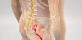
Uterine fibroids are widespread particularly for females from African descent. Many women suffering from fibroids are not symptomatic which could lead to underestimating the extent of its presence. Uterine fibroids that are symptomatic can cause menstrual disorders, anemia infertility, miscarriages abdominal bulge, pressure signs that affect the bowels and bladder (such as constipation or frequent urination) as well as sometimes issues related to coital. This report describes a woman who was 25 years old African American woman with uterine fibroids that caused lower back pain that extended towards upper extremities of the left together with weakness and paresthesias on the right leg. The diagnosis was the lumbar radiculopathy, pre-pregnancy as well as an incidental discovery of the Tarlov cyst. The pregnancy progressed the sciatic pain stopped during one trimester. Sciatic pain may be the result of a sudden growth in the size of the uterus that is caused by the early pregnancy that is co-existing with fibroids that are large. The sciatic pain might not persist all through the pregnancy since the uterus that is growing with large fibroids can be moved away from the point that is causing nerve compression.
Introduction
Uterine leiomyoma also referred to as uterine fibroid is a benign tumor made of mostly smooth muscle mass . It generally develops during women’s reproductive years. From all the signs that are caused by uterine fibroids bleeding symptoms are among the most prevalent. Pressure signs are the most rare and are typically related to the rectum or bladder rather than the nerves that surround them. There are some cases of postmenopausal females who suffer from uterine fibroids, causing gait disorders, lumbosacral pain and lumbar radioculopathy where a hysterectomy alleviated the symptoms [1-33]. This case report describes the case of a 25-year old African American woman with sciatica caused by an expanding pregnant uterus that was also suffering from fibroid that was then relieved as the pregnancy grew in weight and size.
Case Presentation
The patient was a 25 year old nulliparous African American female with a medical history of uterine fibroids as well as benign microscopic hemoglobinuria was diagnosed with a gradual onset of lower back pain that extended into her left lower extremity. The pain gradually grew worse as it was associated with paresthesias in upper extremities left and was not alleviated with non-steroidal anti-inflammatory medications. The patient was active sexually and was not taking contraceptives. The patient was experiencing a regular menstrual cycle.
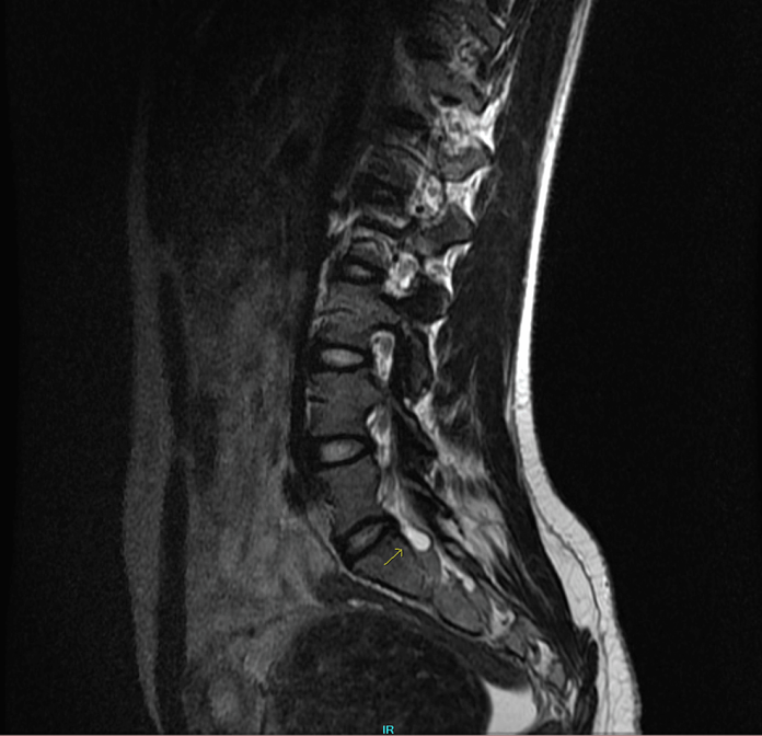
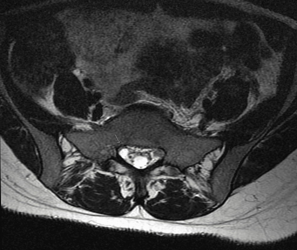
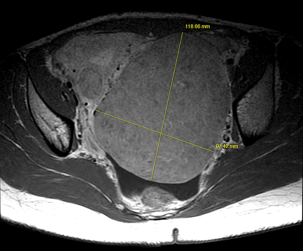
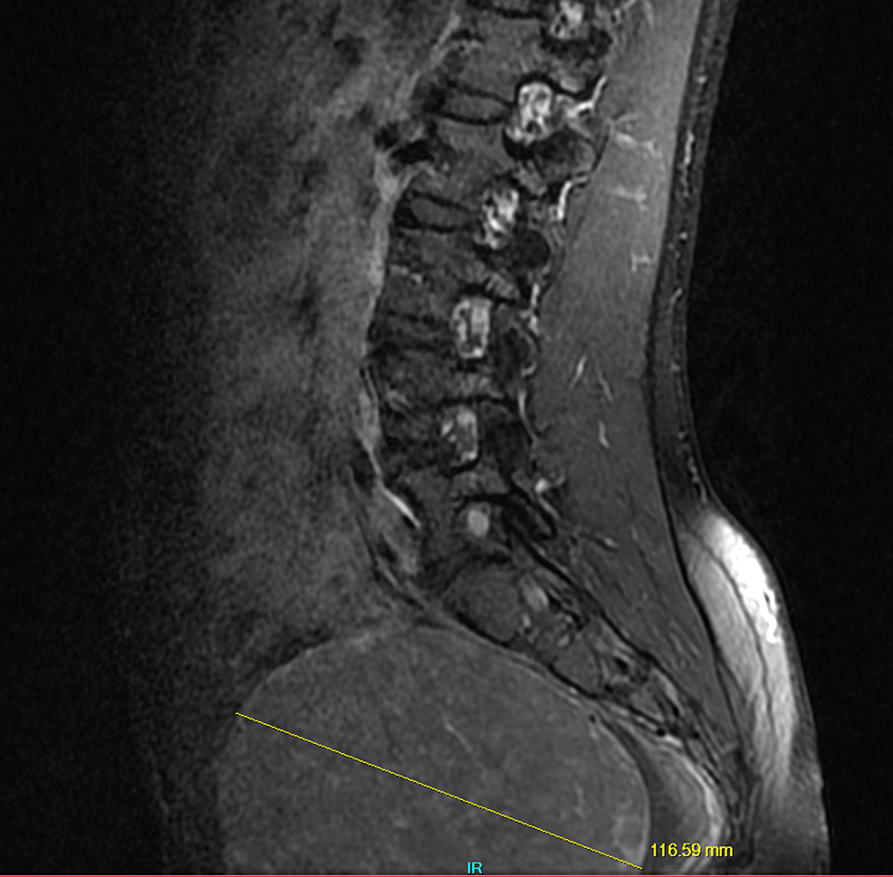
Physical examination revealed that the gluteal and lumbar regions were tender. The range of motion of the left hip was not normal and an examination of the straight leg showed positive results on the left (flexion of the hip when knees were extended caused discomfort). Muscle strength, feeling, and reflexes were all normal. It was determined that the patient suffers as having radiculopathy. She was and was prescribed tramadol 50 mg, to be taken three times daily and scheduled for Magnetic resonance imaging (MRI) of the lumbar spine with or without contrast. The MRI revealed no disc protrusion, or stenosis, however, a small Tarlov cyst was seen at the L5-S1 vertebra to the left (Figures 1 and 2). It was also noted that the MRI scan also revealed an enormous soft tissue pelvic mass of approximately 12 10 cm 12 cm, which is likely in line with her previous history with fibroid (Figures 3 4, 3).
Five days after, a follow-up visit with the patient found the absence of pain relief with tramadol, and the patient was complaining of increasing symptoms, including weakening of the lower extremity of her left. The patient also expressed concern over a missed period of 3 days. The urine test was positive for pregnancy test. The patient was thin and had a the appearance of a prominent lower abdomen upon physical examination. When the patient was examined there was tenderness within both the pelvic region of the left as well as the left hip area. Also, there was a decreased mobility of the left hip when it was flexed. A vaginal and abdominal ultrasound revealed no sac of gestation within the uterus. The gestational age was 4 weeks and three days , based on her menstrual cycle and beta human gonadotropin (hCG) was recorded as 4,800 IU/L. Based on the results above, this patient was identified as having unnatural pregnancy (suspected an ectopic pregnancy) and was scheduled for diagnostic laparoscopy.
The operatively performed scan revealed an extensive, roughly 10 cm fibroid that extended across the entire surface of the uterus up to the left side wall of the pelvis. The pregnancy was not evident anywhere within the pelvis. The patient did well after surgery, however, she was still suffering from sciatica and was prescribed Oxycodone, which helped with some symptoms. It is vital to know that the patient was suffering from the first intrauterine pregnancies that was difficult to see probably because of the technical challenges of ultrasound and fibroids. The subsequent follow-up using an abdominal ultrasound showed an intrauterine pregnancy at 5 weeks and 5 days gestation. In seven weeks into pregnancy the patient’s sciatic pain eased without the need for further pain medications.
Discussion
Sciatica is not a common issue during pregnancy. When pregnant relaxing hormone relaxin loosens ligaments, which causes an alteration in the body’s centre of gravity. This could result in irritation or a pinching of the sciatic nerve [44. Apart from the fibroids and the relaxin hormone, it is possible that it may have contributed to the patient’s sciatica due to its levels being most high in the first trimester pregnancy. While sciatica may occur at any time but it’s more likely to occur during the third trimester because the baby’s weight could cause pressure on the nerve [44.
The accidental discovery of an Tarlov cyst in the patient’s MRI scan might be a cause for Sciatic pain. Tarlov cysts are filled with fluid nerve root sacs that are usually unaffected and may be found in the course of MRI scans. In rare instances cysts can expand and exert pressure on the affected nerve roots, causing Sciatic discomfort, weakness and less sensation in the lower extremity affected. A spontaneous removal from Tarlov the cyst as well as sciatic pain may occur, and is an explanation the reason why her symptoms disappeared without surgical intervention. However the MRI confirmed it was evident that there was a Tarlov cyst was small and was situated on the left aspect (Figure 2) and her symptoms were located on the left side. Due to the size and location of the cyst it’s an unlikely reason for the sciatic pain the patient was suffering from on her left.
The uterine fibroid’s large size seemed more likely to be based on the location of it, as seen by the MRI scan, and also intraoperatively. It was located across the posterior wall of the uterus and into the pelvic floor on left and corresponding to the location of nerve compression to the left (Figure 3). The cases of fibroids with large size causing gait disorders and radiculopathy were resolved with the procedure of hysterectomy, though the cases occurred in post-menopausal and perimenopausal women [1-33]. As menopausal cycles progress, the uterine fibroids can shrink in severity, easing symptoms, but occasionally, fibroids are seen in this age group particularly within African Americans [6]. In a study on a cohort of women by Templeman and colleagues. The second instance of uterine fibroids was observed among women who were 50-54 years old [77. However, in the instance of this woman the symptoms diminished in the first trimester when the baby grew in size. In the course of pregnancy, the uterus expands and is released of the pelvis after 12 weeks gestation. Remember that the size of the uterus did not match the anticipated size at the time of each gestational age due to coexisting fibroids. The patient’s uterus was visible prior to 12 weeks gestation. Additionally the uterine fibroids are known to expand by size in pregnancy due to of the increased sex hormones in the mother which are produced prior to 11-14 weeks gestation [99. It is possible that the rapid growth of the fibroid masses in the first trimester due to the growing uterus increased the pressure upon the sciatic nerve and the fibroid swell was moved due to the growing uterus outside of pelvic floor, thus decreasing pressure on the sciatic nerve.
Pregnancies that have complications with fibroids are usually based in the amount and position of the fibrid. A few adverse obstetric complications which are listed in order of frequency includes cesarean sections and malpresentation, labor dystocia postpartum hemorrhage and peripartum hysterectomy as well as preterm labor and birth [1010. Sciatica is a rare sign of fibroid, and is more common during pregnancy. While the pain eased in the first trimester it’s not uncommon to expect a recurrence later in the pregnancy or even in the puerperium because it shrinks back to its pre-pregnancy size, which is about the same size when it was at the beginning of the sciatic pain. In the case of this patient the pain did not recur in the course of pregnancy. However, after the birth she was afflicted with red swelling of the fibroid and bleeding during the third trimester. She was preterm birth and delivered after 35 weeks of gestation, vaginally. The possibility of a repeat occurrence of sciatica due to fibroid during pregnancy is not unlikely since it will depend on a few aspects. The likelihood of sciatica returning could be based on the uterus’s the position throughout the pregnancy as well as puerperium. Additionally it is possible that diminution in diameter of the fibroid during puerperium may be another reason. Most fibroids don’t changes during puerperium; however, approximately 7.8 percent of them will shrink in size by as much as 10 percent [11,12The volume of the fibroid can decrease by up to 10% [11,12]. A decrease in the volume of fibroids and changes in the position of the uterus may have decreased the chance of the patient’s sciatica returning post-partum or prenatally.
Conclusions
A detailed obstetrical or the gynecological history must be taken into consideration when a woman presents with sciatic pain. further investigation and examination should be conducted on the basis of historical findings. If a patient is suffering from pregnant early and an history of fibroids, presenting in sciatic pain you should think about conservative treatments like analgesic management and close monitoring following the ruling out any other possible reasons.

We understand how important it is to choose a chiropractor that is right for you. It is our belief that educating our patients is a very important part of the success we see in our offices.

