https://www.fapjunk.com https://pornohit.net london escort london escorts buy instagram followers buy tiktok followers Ankara Escort Cialis Cialis 20 Mg

Ask the doctor about flu shots, vitamins B12 Scoliosis. WDU

0
Ask the doctor about flu shots, vitamins B12 Scoliosis. WDU

SOUTH BEND Ind. (WNDU) – – Dr. Bob Cassady from the South Bend Clinic joins us each Thursday on the 16 News Now at noon to answer any medical concerns you may have.


Answer # 1 (from Debra): “How long after getting COVID should one be waiting to get an influenza shot?”

DR. BOB: The health care community has plenty of experience in administering more than one vaccination at once.

If we offer vaccinations to babies usually, they get several shots that include vaccines against more than five distinct organisms.

The CDC guidelines recommend the administration of COVID vaccines regardless use of any other vaccinations. Therefore, the consensus is that you can receive flu vaccine as well as COVID vaccine in the same day.

Since COVID vaccines were created relatively recently, and there’s no quick and easy date when you have to take the COVID vaccine or the flu vaccination, it may be sensible to spread the vaccines out over a week or two.

If you suspect that you will not be able to follow up you might want to for you to call them that next day.


Question 2 (from Shauna): “My friend has started taking B12 vitamin supplements daily and has shed a significant amount of weight. Could this be a coincidence or can these pills help in losing weight? ?”

DR. BOB: Vitamin B12 is an extremely important vitamin for our well-being.

For those who eat an extensive diet that includes grains, fruits, and animal products, deficiencies are rare. I haven’t heard about Vitamin B12 supplementation for those who do not have B12 deficiency, which can cause weight loss.

It’s possible that if someone has a deficiency and they begin taking B12 that they’ll feel more comfortable, which can aid them in exercising and eating more in order to lose weight.

However, I wouldn’t typically recommend taking B12 for solely the aim of losing weight.


Answer 3. (from Erin): I have scoliosis . I also have the sciatic nerve in my right leg. Are they related? and is there something you can try to assist in reducing the discomfort?

DR. BOB Scoliosis refers to an unusual curvature in the side of the spine. It is commonplace and ranges in severity.

The majority of people suffer from minor curvature. However, some individuals may suffer from severe or moderate cases. Scoliosis may increase the chance of having chronic back pain , or other issues like sciatica.

Most people should treat this condition in the same way as in the case of not having the condition of scoliosis. The treatment for sciatica is generally the combination of physical therapy and anti-inflammatory medications.

If someone was suffering from severe Scoliosis, it’s possible there are different options for surgery that could be considered for treating back discomfort.

The most common cause of lower Back Pain, say Doctors You Eat This Not That – Eat This, Not That

0

The back pain can be a frequent problem that can become persistent and even painful. There are a variety of kinds of back pain, and lumbago is an umbrella term used to describe lower back symptoms of pain. It is experienced in the legs from time to time. Based on the International Association for the Study of Pain, “Low back pain is a widespread worldwide issue. The prevalence for low back discomfort (LBP) for 2017 is reported to be around 7.5 percent of the population, or approximately 577.0 millions.” Consume This, Not That! Health spoke to Dr. Bayo Curry-Winchell Urgent Care medical director and physician at Carbon Health, and Saint Mary’s Hospital who gave us everything you need to know about the condition and the symptoms that show that you are suffering from it. Learn more about it and to protect your well-being and that of your loved ones, don’t overlook these signs you’ve had COVID.


Shutterstock


Dr. Curry-Winchell explains, “One moment you’re making dinner, and then the next thing you know, you’re not able to stand up from the sofa. The sudden appearance of not being able stand in a straight line or even walk the typical initial indication of the condition known as lumbago. It can last from several days, or several weeks, or even months. ”


Shutterstock


Dr. Curry-Winchell states, “Leg pain can also be a sign that indicates lower back discomfort due to the related pain that is caused by the shortening of muscles as well as the pressure on nerves on the back.” back. The length of the pain can be varying based on the severity that there is nerve impingement. ”


Shutterstock


Dr. Curry-Winchell describes, “You may experience tightness and soreness or pain in your hips due to the tightening of your muscles in your lower back. It could last for several days, or some weeks or even months.”


Shutterstock

“Lumbago is described as pain in the back, particularly in your lower back,” says Dr. Curry-Winchell. “The discomfort can be caused by many circumstances, but it is most commonly caused by regular activities such as changing your clothes and off, lifting your child up or household chores, such as cutting the lawn or filling the dishwasher. The muscles in your back getting tighter as if were being attacked. This is your body’s reaction to guard your spine from injury. The tightening, which is sometimes referred to as a muscle shortening, can be associated with inability to sit, stand or walk. If you attempt to sit down, your muscles become shorter (tightened) and causes pain immediately.” 6254a4d1642c605c54bf1cab17d50f1e


Shutterstock


Dr. Curry-Winchell explains, “It can be debilitating. The muscles in your back allow you to remain active and permit small and large movements to happen. Everybody feels back discomfort in a different way, but it’s often caused by severe pain that affects your ability to sit and stand. If you notice a an inability to control you bowels, fever or tingling/numbness , it could become a medical issue. it’s crucial to seek treatment as quickly as is possible. ”


Shutterstock


“Lumbago (lower back pain) can affect multiple parts of your body including your hips and legs, making it difficult to pinpoint the root of the pain.” Dr. Curry-Winchell explains.


Shutterstock


Dr. Curry-Winchell advises, “Staying active by participating in moderate or low intensity workouts, strength training using good body mechanics when you movements as well as the lifting of (heavy and light) objects, and maintaining the right diet and weight could help. The greater the chance that your lower back muscles will have to perform regular movement that incorporates proper physique mechanics and a healthy balance of standing and sitting and engaging in exercises that improve your strength and endurance of your core muscles will lower the risk of the aches and pains of lumbago.”

Heather Newgen

Heather Newgen has two decades of experience in writing and reporting about fitness, health entertainment, travel and other topics. Heather is currently working as a freelancer for various magazines. Find out more about Heather

Help is available to back neck and shoulder pain Health The Post Intelligencer

0
Help is available to back neck and shoulder pain Health The Post Intelligencer

Paris Post Intelligencer

How Effective is Chiropractic Treatment?

0

Chiropractic care is a common treatment for various health problems, including back and neck pain. It can also be used to treat soft tissue and joint pain. Other common ailments treated by chiropractors include headaches, allergies, and fibromyalgia. It is a safe and effective way to treat these ailments.

Read More About How Effective is Chiropractic Treatment

american chiropractors second image

More Things To Know About How Effective is Chiropractic Treatment

What is the Success Rate of Chiropractic Treatment?

content machine american chiropractors photos a

Chiropractic treatment has been used to help people with a variety of issues, from chronic back pain to ear infections and colic. Most people seeking chiropractic treatment are middle-aged married men or women with low back pain. The most common complaint is low-back pain, but chiropractic treatments can also help with neck pain and headache.

The first large study of chiropractic use in the United States came out in 1991. The second, more recent RAND study, conducted in 1998, expanded and updated the results. It looked at records from randomly selected chiropractors in the United States and Canada. In both studies, chiropractic treatments reduced low-back pain.

The study also looked at complication rates and the effectiveness of chiropractic treatment for chronic low-back pain. It also measured how many manipulations were necessary for maximum response and its cost-effectiveness. Although this study had limitations, the results indicate that chiropractic care may be a viable treatment for many people suffering from low-back pain.

How Long Do the Benefits of a Chiropractic Adjustment Last?

The benefits of chiropractic treatments will vary depending on the condition. If you have been suffering from chronic pain for years, you may need a longer treatment period. However, if you have just experienced an injury or pain, you may only need a few treatments. This is because your pain hasn’t happened overnight.

A chiropractic adjustment is usually painless, although some patients may experience minor discomfort afterward. This is because the adjustments are meant to treat the problem and prevent it from recurring. It is important to see a chiropractor on a regular basis if you have ongoing back pain.

The benefits of chiropractic adjustments can last for several weeks or months. However, these benefits can also last longer if you follow the prescribed course of treatment. In addition, many chiropractors prescribe several adjustments in a row.

How Often Should You See a Chiropractor?

There are a variety of factors to consider when determining how often you should visit a chiropractor. The number of visits you need will depend on the nature of your condition and how severe it is. You may need several visits to get relief and prevent the pain from recurring. Regardless of the condition, it is important to get regular chiropractic treatment to help keep your body healthy and functioning at its best.

A chiropractic visit can last anywhere from 45 minutes to an hour. In most cases, a visit is shorter than this. During your visit, your chiropractor will assess your condition and recommend a treatment plan that works for you. Some treatment plans may require multiple visits within a few weeks to treat your condition.

Is There Any Evidence That Chiropractic Care Works?

Chiropractic care involves adjusting your body’s spine and joints to relieve pain and facilitate the body’s natural healing process. It can be effective for a variety of health conditions, including back pain and headaches. Most research involving chiropractic care is basic and is performed on a small number of subjects. Generally, this type of research looks at changes in the body in a controlled environment. Some of these studies have examined people with chronic, acute, and subacute lower back pain. If you were an evidence-based chiropractor, you would promote these results to patients.

While there is a high level of uncertainty about whether chiropractic care can effectively cure back pain, some studies indicate that it can relieve pain. In fact, it has been estimated that 22 million Americans seek chiropractic treatment each year, including 7.7 million people who suffer from back pain. Chiropractors perform spinal manipulation to restore the range of motion to joints that have been restricted by disease or trauma.

How Long Does It Take to Realign Your Spine?

After chiropractic treatment, you can expect a significant improvement in your spine alignment. In some cases, you can feel results in as little as a week. However, the number of visits will decrease over time as your body regains its balance and strength. For most patients, a few treatments are enough to see a noticeable improvement in their alignment.

A chiropractor uses manual manipulation to realign your spine. This involves stretching, thrusting, and applying pressure to the spine. The chiropractor will first assess your current condition and determine the specific technique to use. Then, he or she will use specific controlled forces according to the specific pain points in your spine. You will feel only a moderate amount of pressure during a chiropractic adjustment.

Headaches: A common, challenging comorbidity Psychiatric Times

0
Headaches: A common, challenging comorbidity Psychiatric Times


TALE from the clinic


-Series Editor Nidal Moukaddam, MD, PhD

In this episode of Stories From the Clinic: The Art of Psychiatry, we look into a common neuropsychiatry-related comorbidity: headaches. This condition of the neuropsychiatric system is a frequent comorbidity among various psychiatric diagnoses and is often difficult to manage due to its significant connection with somatization.


Case Study

“Mr Wright” is a 37-year-old male with no medical comorbidities and has been living in his home in the United States for more than 10 years. He was seen by primary care for a 4 month background of headaches that weren’t responding to nonsteroidal anti-inflammatory medications available over-the-counter (NSAIDs). The patient is prescribed amitriptyline 25 mg daily at bedtime and is then referred to the department of psychiatry for “anxiety” with no other notes about the signs of anxiety. The patient is afraid that they will label him “crazy” as well as unhappy because his struggles are not being treated with seriousness.

He is physically healthy with excellent grooming habits, however the man is a bit hesitant in the psychiatric examination. He says he’s lived with his spouse and child and works in the restaurant. There are teenaged children that are not living with him, however the reports say that he has a great relationship with them.

In describing his headaches When describing his headaches, Mr. Wright states that they last throughout the day and cover areas surrounding his bilateral temples as well as the occipital zone but the pain can be transferred between these regions. He’s tried lying down for a rest however the relief from discomfort is only temporary. He’s not able to focus on driving, and has had to miss several working days because he did not think he was able to pay attention when driving. He is able to stretch his neck at several different positions in our conversation to alleviate the pain. Along with a lot of eye blinking that he is unaware of. He denies that a premonitory feeling was which occurred before his motions.

After multiple inquiries about the causes of his stress at the time when his headaches began, Mr. Wright can recall the incident that occurred one week prior to their onset The incident occurred when he was assisting his sister in his home following an argument between her and husband. His brother-in law had held him in a gun-point situation outside of his home. He fled to security, and his brother-in-law later went away. The sister who was with him has now gone and the patient believes that the stress of an unkind relationship with his sister continues to be a problem for his sister as well as the other members of the family. He remembers an incident similar to this about 20 years ago, when the patient was chased by a family member from another in his country of origin. At the time, Mr. Wright felt there was a danger to his safety, and needed to escape towards safety. Since the time, he’s been a nervous person generally. He has not received any treatment for mental health (including therapy).

He is not experiencing the typical signs of Posttraumatic Stress Disorder (PTSD) such as flashbacks, nightmares, and avoiding the arousal of heightened levels.

A review of the chart shows that Mr. Wright was examined in the emergency room twice following his visit to his primary physician. One time the patient complained of severe chest pain. Negative EKG as well as cardiac enzymes resulted in an admission to the hospital. In the next instance, he was screened for sexual contact that was not protected by an unidentified individual. Mr. Wright was hesitant to discuss any of these incidents of his interactions with medical professionals, and the psychiatrist opted not to speak about them.

Amitriptyline was recommended to him at 25 mg with his main doctor two weeks ago. He is taking it for the last two weeks when that he’s unable to fall asleep. It has been helpful to him however, he is unable to get any relief from headaches. He’s unable to express the connection between a potentially life-threatening incident and the beginning in his headaches.

After the consultation, both physician and patient are in agreement that the amitriptyline treatment should be maintained, however the patient needs to use it more often to get the most effective therapeutic results.


Discussion

As per a systematic review of a global burden of disease across the United States, in 2017, the 5 highest incidence rates of neurological diseases included migraines and tension-type headaches.1 Migraines have consistently been among the top 3 neurological diseases in terms of disability-adjusted life years.2 As a testament to how frequently psychiatrists and neurologists see patients with headaches and psychiatric diagnoses, in 2019, the most frequent psychiatric comorbidities in individuals with primary headaches in the United States were anxiety and depression, followed by posttraumatic/stressor and trauma-related stress disorder. The comorbidities mentioned above are present in 25 percent, 23% and 15 percent of the general population according to. 3

It is therefore inevitable that you will encounter patients suffering from both principal and second headaches in the psychiatric clinic. The recognition of headaches is a valuable instrument to assess the health for our patients. The headache can trigger and perpetuating causes of psychiatric disorders and are important in determining the most effective pharmacological treatments.


Biopsychosocial Model

In primary headaches such as migraine or tension headaches there is no reason to believe that there is a nociceptive cause such as hemorrhage, or inflammation. There are many theories that have attempted to explain the relationship between stress and the onset of primary headaches. The results of studies that have examined the connections between stress and primary headaches clear that the main trigger factors for primary headaches are stress and sleep problems. 4 Stress has been associated with the change from chronic migraines into chronic migraines which carry a larger burden of illness. 5 Interestingly repeatedly exposed to trauma regardless of an official diagnosis of PTSD, is more prevalent in people who suffer from chronic migraine headaches when compared to adults who do not suffer from migraines.

Biopsychosocial variables are believed to play a role in the perception of headaches. Transactional models suggest that the gap between the perception of threat and the ability to face the threat may be applied to the subjective feeling of pain that occurs in migraines and to any other type of pain syndrome. 6 Among personality traits, the locus of control is perceived as external versus internal is a predictor for better outcome for headache.

In the COVID-19 epidemic, the significance of a sudden shift in the mental health due to the shutdown in people with prior migraines is being investigated. Contrary to the belief that headaches are likely to worsen, findings from at the very least one study indicate improvements. The results show a decrease in frequency and intensity of migraine among adolescents during the closure within Italy from 2020. 7 It is possible to infer that the decrease in social and academic pressures was beneficial in this improvement. It is also important to highlight the importance of psychosocial treatment in patients suffering from migraines. Additionally, it is fascinating to know whether there’s an increase in migraine presentations to psychiatrists and neurologists because of the constant anxiety caused by the pandemic, which is over and beyond migraines in patients who suffer from the neuropsychiatric consequences of COVID-19 itself.


of Neuropsychiatry of Headache

Migraine headaches have a bidirectional connection with a range of mental illnesses, with the most significant correlation is with depression anxiety, depression, and bipolar disorder. There is evidence of a shared genetic predisposition, aswell being structural and functional changes in brain regions that control pain such as the amygdala the anterior cingulate cortex, and the periaqueductal grey in the affective and migraine disorders. Additionally, they share problems within the HPA axis, as well as similar mechanisms for action for treatments.

Central sensitization is commonly used to explain conditions that cause pain which include migraine. People suffering from chronic migraines (as contrasted with episodic migraines) suffer more from somatic complaints. The significance of pain sensitization in episodic migraines is also being studied in the context of episodic migraines. A recent study suggests that evoked pain sensitization may be a useful diagnostic indicator for intermittent migraines. 8

Serotonin, norepinephrine and norepinephrine, as well as the GABAergic system have have been linked to the cause of psychiatric and primary headaches on the neurotransmitters level. Therefore, the evidence to use serotonin and norepinephrine Reuptake inhibitors (SNRIs) and tricyclic antidepressants (TCAs) which are targeted at these receptors to prevention-based treatment of migraines remains solid.


Pearls in Management

Treating psychiatric issues in patients who are seeking treatment for headaches could provide a distinct advantage when it comes in addressing the issue of improving mental health. When setting goals for treatment it could be beneficial to explain to the patient the benefits of SNRIs and TCAs in the management of headaches and anxiety and mood issues. It can help patients communicate their needs for mental health even in the face of stigma. Sometimes, offering such an SNRI as well as TCA as a headache medication is a great way to improve compliance to treatment for patients who struggle to communicate their feelings.

One of the biggest difficulties when it comes to managing headaches is that introducing the use of SNRIs or TCAs to treat headaches can be a source of headaches. Careful consideration of the time frame for headaches caused by medication can help in the separation of symptoms that are superimposed. Clinicians might benefit from considering the withdrawal of medication as a possible cause for headache exacerbation in treating patients suffering from psychiatric disorders with comorbid headaches and having a history of nonadherence. Venlafaxine is prominent in this regard due to its withdrawal symptoms.

Researchers of a variety of studies have not been able to prove the effectiveness of making use of selective serotonin reuptake inhibitors (SSRIs) to prevent migraine Relapses. One of the most researched SSRIs are fluoxetine. It to the present, has inconsistent evidence of its effectiveness for primary headaches. 9 Other clinical trials of sertraline as well as citalopram have not shown any advantages when compared to TCA as well as placebo. So, if a person who has previously received treatment using SSRIs is put on TCAs to treat headaches and headaches, it is necessary be mindful of the potential advantages and risks when switching from monotherapy using the use of a TCA or continuing treatment with both medication. If the patient has reported positive clinical results, they can be treated using low dosages of both medications and be monitored for any adverse reactions. Patients may be taught about the early signs of serotonin syndrome when being treated in an outpatient setting.

Furthermore, many physicians have concerns about the likelihood of serotonin-related serotonin disorder in those taking antidepressants that are prescribed triptans to prevent migraines. migraines. While there was a black-box warning about this interaction, findings from an electronic health record study conducted in 2018 did not reveal any connection between serotonin syndrome and co-use of SSRIs as well as SNRIs or triptans. 10

In patients who suffer from drug-overuse headaches there is a higher incidence of psychiatric comorbidities may be more common. 11 Some mention the similarity with the use of compulsive medications for headaches and those with obsessive compulsive disorder. In this regard, the addition of the behavioral treatment for this segment of patients can improve results for headaches.

It could be beneficial that neurologists prescribe regular psychotherapy for patients suffering from chronic migraines. Forecasting models and behavioral interventions that incorporate stress factors are effective in the management of migraine. The behavioral treatments that are proven effective for migraine control include: stress management as well as relaxing therapy, therapy as well as mindfulness-based treatments, biofeedback as well as acceptance and commitment therapy. Researchers of certain studies note that even though the intensity or frequency of headaches decreases however, there is no improvement in the physiological markers that cause headaches like tension in muscles. In a groundbreaking study on electromyographic biofeedback participants perceived the source of their headaches as having an internal source of control, and also as being more self-efficacious (ie that they are capable of altering their headaches) in the aftermath of the treatment. 12


Final Thoughts

In the final analysis, when dealing with this diverse patient population, it’s important to avoid labeling or stigmatization. Patients are typically worried about physical ailments that are ignored or being advised that “the symptoms are just in the head.” A an appropriate therapeutic relationship will lead to discussion of treatment options that are open and opens the way for effective management of illness.

“Dr Shamim has been psychiatrist from Houston, Texas.


References

1. GBD 2017 US Neurological Disorders Collaborators; Feigin VL, Vos T, Alahdab F, et al. A Study of the Burden and Impact of Neurological Disorders across the US Between 1990 and 2017 A Global Burden of Disease Study. JAMA Neurol. 2021;78(2):165-176.

2. GBD 2019 Injuries and Diseases Collaborators. The global burden for 369 ailments and injuries in 204 nations and territories between 1990 and 2019 A systematic analysis for The Global Burden of Disease Study 2019. Lancet. 2020;396(10258):1204-1222.

3. Caponnetto V, Deodato M, Robotti M, et al; European Headache Federation School of Advanced Studies (EHF-SAS). Primary headache disorder-related comorbidities Review of the literature and meta-analysis. J Headache Pain. 2021;22(1):71.

4. Yokoyama M, Yokoyama T, Funazu K, et al. The relationship between stress and headache alcohol consumption as well as exercise, sleep and other health issues in the Japanese population. J Headache Pain. 2009;10(3):177-185.

5. Xu J, Kong F, Buse DC. Predictors of episodic migraine’s transformation to chronic migraine: A comprehensive review of and meta-analysis cohort studies of observation. Cephalalgia. 2020;40(5):503-516.

6. Lazarus RS, Folkman S. Stress, Appraisal, and Coping. Springer Publishing Company; 1984.

7. Dallavalle G, Pezzotti E, Provenzi L, et al. The symptoms of migraine improve after the COVID-19 lockdown an array of adolescents and children. Front Neurol. 2020;11:579047.

8. De la Coba P, Bruehl S, Del Paso GAR. Slowly repeated provoked pain (SREP) as an indicator of central sensitization for migraine patients with episodic attacks. Sci Rep. 2021;11(1):4582.

9. Silberstein SD, Holland S, Freitag F, et and Quality Standards Subcommittee of the American Academy of Neurology and the American Headache Society. A new guideline based on evidence: pharmacologic treatment of episodic headache prevention for adults Report by the Quality Standards Subcommittee of the American Academy of Neurology and the American Headache Society. Neurology. 2012;78(17):1337-1345.

10. Orlova orlova Y, Rizzoli P, Loder E. Association of coprescription of triptan antimigraine medications and selective serotonin reuptake inhibitors or selective norepinephrine-reuptake inhibitors of antidepressants for serotonin-related syndrome. JAMA Neurol. 2018;75(5):566-572.

11. Kristoffersen E, Straand J Russell MB, Lundqvist C. Disabilities anxiety and depression among patients suffering from headaches caused by medication in primary treatment The BIMOH study. Eur J Neurol. 2016;23(suppl 1):28-35.

12. Holroyd KA, Penzien DB, Hursey KG, et al. Mechanisms of change involved in EMG Biofeedback Training: changes in the brain that cause improvement for tension headache. J Consult Clin Psychol. 1984;52(6):1039-1053.

Are Mike Tyson OK? The former Heavyweight Champion spoke about Death Recently. Distractify

0
Are Mike Tyson OK? The former Heavyweight Champion spoke about Death Recently. Distractify

On the show, Mike spoke with therapist Sean McFarland, who specializes in addiction and trauma. In their conversation, Mike gave a pretty shocking statement about his passing. “We’re all going to die some day, no doubt,” he said to Sean and added, “Then, when I glance at my reflection, and I see these little marks on my face. I think, ‘Wow!. It means the expiration of my contract is approaching very in the near future. ‘”

Lifetime Gator Discovers Back Pain Relief Through UF Health – UF Health

0

If Bill Belote could choose one spot to be in then it would be on his tennis courts. He’s played for all his life, and was part of the University of Florida men’s team during his time in college. But, two years later, after the back pain began and he was forced to retire from tennis courts came to stop. It was a transition from active to being unable to enjoy the activities that he enjoyed, like gardening, and obviously playing tennis. The sciatica pain spread through his legs, which caused an increase in impermanence.

Bill was aware that he needed to find relief. He went for images as well as an MRI at another hospital. There, the doctor told him that he’d require a long, complex procedure that would cause trauma to the back muscles, force the wearer to wear a back brace, and take months to recover. Bill wanted to seek an opinion from a different doctor, but was not certain where to take his next step; he wanted to ensure that there was a doctor that who he could believe in. At the same time that he was at an UF tennis game, and the match was played next to a close friend who was a student of Adam Polifka, MD, an associate professor at the UF Department of Neurosurgery. After hearing more about the Dr. Polifka and UF Health from his friend and then, it all was in place.

“All for the “Gators”

As a child within Jacksonville, Bill had been an avid Gator for the longest time his memory could allow. His mother was determined to give him all she could. While caring for and working Bill in the role of a single mother, she also earned her doctorate and master’s degrees in education. When he was a child playing in summer sports at the P.K. Yonge Developmental Research School, it was his chance to meet with a coach from his UF football team. He also had the chance to see matches from the benches starting at the age of 10 until when his high school graduation. He knew that UF was the place he wanted to for college and was certain he wanted to play football for the Gators and was successful in both.

Discovering UF Health

With his long-standing love and history with his beloved University of Florida, Bill knew that attending UF Health was the right decision for him. was unsure why he didn’t enroll at UF Health sooner. Following that fateful game of tennis, Bill sent his X-rays as well as other images for the doctor Dr. Polifka, who knew that the faster he completed surgery on Bill the higher the quality of his life was going to be.

Dr. Polifka’s treatment differed from the advice Bill had been told by the first doctor he visited. Doctor. Polifka used a minimally intensive lateral approach to minimize injury on his back muscles and to speed up the healing time. Bill wouldn’t even have the use of an back brace. In the spinal procedure the doctor. Polifka used a 3D titanium cage that was printed to relax the nerves that were squeezed by the sliding in the L4 vertebrae over the L5 vertebrae. He then joined both the L4 and L5 vertebrae.

After the operation, Bill felt instant relief — a moment that changed his life that he’ll never forget. He was aware that there would be recovery time however he knew that the first step was to being back to playing tennis and gardening. He was hospitalized for 3 days, 2 nights at hospital. During that period, he was grateful for the staff members residents, nurses, and other staff members who assisted him throughout his hospital stay.

“I cannot say thank you to the Dr. Polifka and his team enough for all they did for me. If someone has to undergo significant surgery they may experience fears and anxieties however, know that the skilled doctors in UF Health can fix your issues with the least amount of risk.”

With the help of Doctor. Polifka and UF Health, Bill is back doing his passions: playing tennis and working in his yard and leading a pain-free existence.

“As to me the Dr. Polifka is a miracle practitioner. He brought an extremely debilitating and painful life back into normal.”

Migraine headaches – South Platte Sentinel

0
Migraine headaches - South Platte Sentinel
Mark McDonald

It is believed that migraine headaches are a particular kind of headache that a lot of sufferers experience throughout their lives. The condition affects 1 in 7 people, mostly between the ages 15 to 55, and women are three times more likely migraines than males. Within the United States, 39 million suffer from migraine, which makes migraine one of the top debilitating ailments in the world.

Migraine is a condition of the nervous system that can trigger intense pain in one side or the other of the head that lasts for up days to up to 72. It’s a type of recurring of headache. However, unlike other headaches the symptoms typically is accompanied by pulsing or throbbing headaches, tension in the temples or behind one eye or the ear as well as sensitivity to light and sound.

There are two kinds of migraine: migraine that has or without aura. Aura is a stage of migraine attacks where patients may experience flashes of illumination, see blind spots or feel tingling in their hands or on their faces. A few sufferers experience migraines one or two times a month, while other suffer migraines for 15 or more days per month. While everyone suffers from migraine differently but the effect is usually disrupts their daily life. The discomfort, nausea as well as fatigue and sensitivity cause a lot of difficulty in functioning and can disrupt daily life including school, work and social activities.

One in five patients experiences aura. The aura is the initial phase of migraine that may begin in as little as 24 hours before an attack. It is also known as prodrome and is characterized by eating cravings, mood swings in the form of uncontrollable yawning or difficulty concentration. The triggers for migraine can be anxiety, stress, too little or excessive sleeping, exhaustion from physical causes, and menstrual changes and hormonal changes. Food triggers can include alcohol, tobacco, caffeine, processed and cured meats in addition to dehydration, skipped meals and a lack of. Influences from the environment include weather fluctuations and flashing lights, as well as bright or bright and strong odors. Certain medications like migraine medications (interestingly) when used excessively can trigger migraine.

Migraine disorders may also trigger postdrome syndrome, which occurs after migraine headaches. can occur following migraine headaches. Postdrome syndrome may be similar to symptoms seen in prodrome.

As you will see, a migraine can be very debilitating and very particular type of headache that should not get confused by tension headaches or cervical-related headaches (neck-related). If you suffer from a migraine that is real There are treatment options which can aid. Researchers believe that a reason for migraine pain could be linked to a protein known as CGRP or calcitonin gene related peptide. The CGRPs trigger nerve inflammation that triggers the migraine attack.

As you are aware, when you suffer from migraine this isn’t something you’d want to like to see your most hated enemy. For more details on migraine and how it can be treated please do not hesitate to reach out to your doctor for a free consultation. We appreciate your time.

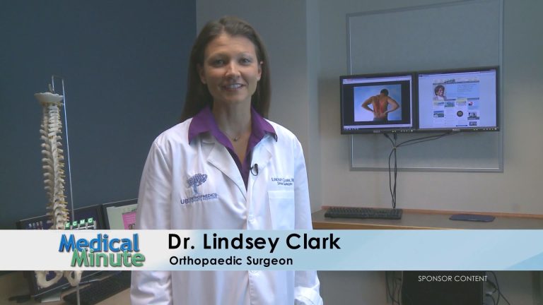
EMC Medical Minute: Dr. Clark – What is Sciatica 09/12/22 Erie County Medical Center

0
EMC Medical Minute: Dr. Clark - What is Sciatica 09/12/22 Erie County Medical Center

ECMC Medical Minute – Dr. Clark – What is Sciatica? – 09/12/22

Join us every Monday morning from 6:30 AM and 6:30 AM on WGRZ-TV, Channel 2 Daybreak, for our ECMC Minute. Medical Minute.

We’ll discuss the most recent health-related initiatives at Western New York, around the world and in the United States. You’ll have the opportunity to participate in the conversation and offer suggestions for future episodes. Are you missing an episode? Don’t worry! We’ll replay every episode on Saturday Daybreak from 9:00 AM until 10:00 AM.

Analysis of Chronic Low Back Pain Caused by Lumbar Microinstability Af | JPR – Dove Medical Press

0

Introduction

Lumbar disc herniation (LDH) is the most common cause of sciatica and accounts for the greatest social health burden in terms of disability and work absenteeism.1 Patients may resort to surgical intervention because of severe low back pain and leg pain that affect their quality of life. Although there are some controversies in terms of surgical indications and methods, percutaneous endoscopic transforaminal discectomy (PTED) is an accepted surgical procedure worldwide for patients with a single-level lumbar disc herniation.2 This method can be used to resect herniated intervertebral discs through a very small incision with the advantages of a low risk of soft tissue injury, fast rehabilitation, and preservation of the motion function of the operated segments.3,4 After surgery, the symptoms of the nerve root can be relieved immediately in most patients, and as many as 95% of patients can achieve satisfactory patient-reported outcomes.5

However, some patients are prone to persisting chronic low back pain (CLBP) with visual analogue scale (VAS) scores higher than 3 for one year after PTED, which is even worse than that preoperatively, and affects their postoperative recovery and the early return to normal.6 Theoretically, many factors are involved in chronic low back pain, including inflammation, muscle degeneration, and facet joint fluid, but the major cause may be related to inherent lumbar instability preoperatively or iatrogenic lumbar instability postoperatively.7 In a previous study, Iguchi et al8 found that the critical factor for low back pain was lumbar instability, and the results revealed that patients with a higher degree of lumbar instability had significantly more days with pain and more hospital visits for their symptoms than other patients. Thus, lumbar instability is commonly the first consideration in determining the treatment strategy.

Although there are multiple modalities for diagnosing lumbar instability, the most common diagnostic technique is the use of lateral flexion and extension standing radiographs.9 However, these have several limitations. Patients cannot perform adequate flexion and extension due to serious pain when standing, which causes analgic contraction and altered muscle tone, which indicates that it is a poor method to reveal lumbar instability.10 Recent studies have proposed many theories regarding lumbar motions, including dynamic instability, lumbar dysfunction, and lumbar microinstability (MI).11 Moreover, Landi et al12 defined clinical condition-specific pathoanatomical and clinical characteristics in the vertebral segment of interest without the presence of spondylolisthesis in flexion–extension radiography as indicators of lumbar microinstability, and found a close relationship between lumbar microinstability and adjacent segment disease (ASD). This publication indicated that low back pain might correlate with lumbar microinstability, and this theoretical background can also apply to patients with CLBP after PTED. However, there is a paucity of research using lumbar microinstability to investigate the motion characteristics of the involved segment or surgical outcomes. Thus, the purpose of this study was to assess the radiographic characteristics of patients and evaluate the effects of lumbar microinstability on patient-reported outcomes for single-level LDH patients who underwent PTED.

Methods

Patients

After approval by the Ethics Committee of Zhejiang Hospital, we retrospectively reviewed the medical records of patients who underwent PTED from August 2018 to March 2021 at the hospital. All methods were performed in accordance with relevant guidelines and regulations and informed consent was obtained from all subjects. We followed the Declaration of Helsinki guidelines. Patients enrolled in this study had to meet the following inclusion criteria: (1) patients with PTED due to single-level LDH; (2) patients’VAS scores for leg pain and low back pain were lower than 4 at the one-year follow-up after operation. (3) patients with PTED were followed up on a regular basis for at least a year; Exclude any of the following: (1) obvious vertebral column Canal stenosis, such as intermittent claudication; (2) mobile movement, the width of the adjacent vertebral body at 3 mm, and/or >8% of the affected segment;13 (3) patients with multi-level lumbar disc herniation; (4) patients with spinal fractures; (5) patients with spinal infections; (6) trauma patients; (7) a cancer patient; (8) patients with a history of lumbar spondylosis.

Surgical Technique

All patients received the same surgical treatment. The PTED patient was placed in the prone position under local anesthesia. Through C-arm lateral fluoroscopy, the surgical segment and the puncture needle point were determined. Puncture at 12–14 cm of the incision gap and make a mark. With the needle near the midline of the pedicle, the needle is located on the disc and the posterior edge of the spine under lateral fluoroscopy. A guide wire was used instead of the needle to pass the dilator through the guide wire into the working cannula. Using forceps and a bipolar radiofrequency coagulation device, the herniated disc fragments were removed endoscopically. Pay attention to the space between the lumbar intervertebral disc and the ligamentum flavum, and ensure sufficient relaxation. After surgery, the surgeon determines the following criteria for decompression: the nerve tissue can move on its own, the dura mater and nerve roots beat autonomously (in synchrony with the heartbeat), restore the anatomical position of the nerve tissue, and improve the blood supply to the nerve tissue. The surgeon also needs to ensure that symptoms have subsided.

Evaluation of Baseline Characteristics

Baseline characteristics included patient factors (age, sex, BMI and vertebral level of the operation), basic radiographic parameters on X-rays (Van Akkerveeken’s lines, Hadley’s “S” curve, Ullmann’s line), and CT (osteoarthritic facet degeneration grade and facet tropism) and MRI findings (Pfirrmann grading, modic changes, facet fluid measurement, and muscle fatty degeneration). The visual analogue scale (VAS) scores for leg and low back pain were used to evaluate patient-reported outcomes preoperatively, at 1 month, 3 months, 6 months and 1 year postoperatively. The Oswestry Disability Index (ODI) was used to evaluate patient-reported outcomes preoperatively, at 3 months, 6 months and 1 year postoperatively. CLBP was defined as VAS ≥4 at every point, because VAS ≥4 was defined as moderate pain.

Radiographic Evaluation

All radiographic parameters were performed twice and averaged to reduce the errors by a single spine surgeon, the pathoanatomic alterations in relation to spinal motor unit dysfunction were analysed, and a specific score was given to every kind of alteration. In X-rays, the Van Akkerveeken’s lines12 refer to the distance between the crossing of the two lines passing through the endplates and the posterior wall of the vertebral body. Hadley’s “S” curve12 is an S-shaped line passing through the inferior margin of the transverse process and the lateral margin of the articular mass. An interruption of the S-shaped line means facet subluxation. Ullmann’s line12 is a line passing through the endplate of S1, and the other line is perpendicular to the first line and passes through the sacral promontory. If L5 is beyond the line, the segment is unstable. In addition, the slip distance percentage was measured as the interval between two extended lines of the posterior aspects of the two vertebral bodies with herniation in dynamic X-rays, which were used to evaluate lumbar instability whether or not (Figure 1).

Figure 1 The Van Akkerveeken’s lines were measured as the distance between the crossing of the two lines passing through the endplates and the posterior wall of the vertebral body. “a” is the distance between the crossing of the two lines passing through the endplates and the midpoint of the posterior edge of the lower vertebral body. “b” is the distance between the crossing of the two lines passing through the endplates and the midpoint of the posterior edge of the upper vertebral body (A). Ullmann’s line is a line that passes through the endplate of S1, and the other line is perpendicular to the first line and passes through the sacral promontory (A). Hadley’s “S” curve is an S-shaped line passing through the inferior margin of the transverse process and the lateral margin of the articular mass (B).

On CT, the osteoarthritic facet degeneration grade14 evaluates the grade of osteoarthritic facet degeneration and includes 4 progressive grades. Facet tropism15 was measured as the difference in the angle between the midline and the line passing through the articular rim.

On MRI, Pfirrmann grading16,17 is a method for evaluating intervertebral disc degeneration, which includes 5 progressive degeneration grades that are related to the degree of progressive instability. Modic changes18–20 evaluate the degeneration of the endplates, and include 3 progressive degeneration grades. Facet fluid21 refers to a large (>1.5 mm) facet effusion. Muscle fatty degeneration22 evaluates whether the lumbar multifidus muscle has fatty infiltration.

A score of 0 is assigned if there is no alteration, or a score of 1 is assigned if the specific alteration is found (ie, the presence of facet fluid, muscle fatty degeneration, facet tropism and of the measurements on the X-ray). The score assigned ranges from 0 to 4 depending on the specific alterations, including modic changes, Pfirrmann grading, and osteoarthritic facet degeneration grade (Table 1).

Table 1 Scores for Radiographic Data

The sum of the different scores defines a total score varying from 0 to 14 and indicates the degree of lumbar dysfunction, where a score of 0 indicates a stable lumbar spine and scarce clinical meaning and a score of 14 indicates severe microinstability.

Study Groups

The enrolled patients were divided into three groups: a stable group (Group S), a dysfunctional group (Group D) and a microinstability group (Group M), based on the sum of the different scores obtained in individual examinations. Group S included patients with scores of 0–3, which was defined as a stable lumbar spine, which indicates that the clinical and radiologica, findings indicate dysfunction in its initial stage and that there is little clinical and biomechanical meaning of the findings. Group D included patients with scores of 4–8, which was defined as a dysfunctional lumbar spine, with accompanies moderate clinical and biomechanical meaning of the findings. Group M included patients with scores of 9–14, defined as microinstability of the lumbar spine, which indicates advanced-stage dysfunction with great clinical and biomechanical of the findings.

Statistical Analysis

Two primary analyses were conducted. First, baseline characteristics were compared among Group S, Group D and Group M. Second, the radiographic characteristics were compared among the three groups. All data were analysed using SPSS version 24.0 (SPSS, Chicago, IL, USA). The data were fit to a normal distribution, so the paired t-test was used to analyse the intragroup differences. Analysis of variance (ANOVA) and the chi-square test were used to analyse the differences among these three groups. Moreover, logistic regression was performed to ascertain potential related and independent risk factors for CLBP. A P value <0.05 was considered statistically significant.

Results

Baseline Characteristics

A total of 127 patients who underwent surgical treatment with PTED were included in this study. The average age of these patients was 50.6 years (range 25–87 years). Of the 127 patients, 71 (55.9%) were male, 31 (24.4%) patients were assigned to Group S, 59 (46.5%) patients were assigned to Group D, and 37 (29.1%) patients were assigned to Group M. There was no difference in reporting among the groups in age, sex, or BMI. Their demographic information was recorded (Table 2). L4-5 was the most common surgical level (n = 76, 59.8%), followed by L5-S1 (n = 46, 36.2%) and L3-4 (n = 5, 3.9%). In addition, the follow-up period was not significantly different among Group S (22.4 ± 7.3), Group D (20.8 ± 8.9) and Group M (20.3 ± 10.3).

Table 2 Baseline Characteristics

Evaluation of the Radiographic Characteristics

The measurements from the X-rays are shown in Table 3. In Group M, the Van Akkerveeken’s lines and Hadley’s “S” curves were significantly higher than those in the other two groups (P < 0.01, P = 0.04, respectively), whereas no significant difference was observed in the Ullman lines (P = 0.11). In the CT images, compared to Group S and Group D, Group M demonstrated significantly higher osteoarthritic facet degeneration (0.26 ± 0.44 versus 0.76 ± 0.60 versus 1.54 ± 0.69, respectively) and different facet tropisms (0.29 ± 0.46 versus 0.56 ± 0.50 versus 0.86 ± 0.35, respectively) (Table 4). As shown in Table 5, the greatest degenerative indication was observed in Group M via Pfirrmann grading (2.83 ± 0.44), facet fluid (0.85 ± 0.35), muscle fatty degeneration (0.81 ± 0.40) and modic change (1.08 ± 0.54). The logistic regression analysis results for low back pain (VAS≥4) revealed that muscle fatty degeneration (95% CI, 1.20–8.51, P = 0.02) and facet tropism (95% CI, 1.39–11.37, P = 0.01) were independent risk factors (Table 6)

Table 3 Radiographic Findings in the X-Rays

Table 4 Radiographic Findings in the CT

Table 5 Radiographic Findings in the MRI

Table 6 Logistic Regression Analysis for High Risk Factors for Low Back Pain

Patient-Reported Outcomes

For patient-reported outcomes, preoperative symptoms including VAS for leg pain and low back pain and ODI scores were not significantly different among the three groups, and all patients achieved significant clinical relief after surgery (ODI scores, P < 0.01; VAS of leg pain, P < 0.01; VAS of low back pain, P < 0.001). However, the scores of ODI and VAS for low back pain scores in Group M were significantly higher than those in the other two groups at 1 month (VAS scores for low back pain, 2.6 ± 0.7 vs 3.5 ± 1.2 vs 4.2 ± 1.4, Group S, Group D, Group M, respectively), 3 months (ODI, 18.7 ± 4.39 vs 21.2 ± 4.09 vs 23.3 ± 3.87; VAS scores for low back pain, 2.2 ± 0.7 vs 2.4 ± 1.1 vs 3.9 ± 1.2, Group S, Group D, Group M, respectively), 6 months (ODI, 16.6 ± 3.91 vs 19.4 ± 3.75 vs 21.4 ± 4.46; VAS scores for low back pain, 1.9 ± 0.5 vs 2.2 ± 1.0 vs 3.8 ± 1.2, Group S, Group D, Group My, respectively) and 1 year postoperatively (ODI, 15.4 ± 3.71 vs 17.0 ± 3.93 vs 20.1 ± 4.24; VAS scores for low back pain, 1.8 ± 1.1 vs 2.1 ± 1.3 vs 3.7 ± 1.1, Group S, Group D, Group M, respectively), as shown in Table 7 (P < 0.05). Additionally, the VAS (leg pain) scores were not significantly different among the three groups at different time points after percutaneous endoscopic transforaminal discectomy surgery (1 month, P = 0.14; 3 months, P = 0.09; 6 months, P = 0.15; 1 year, P = 0.08).

Table 7 Comparison of ODI Scores, VAS (Leg Pain) and VAS (Low Back Pain)

Discussion

The CLBP after PTED surgery was associated with an increased incidence of postoperative complications and decreased patient satisfaction.23 Various studies have reported the risk factors for CLBP after PTED, which include BMI, level of surgery, paraspinal muscle degeneration, sex and Modic changes,24,25 but lumbar instability is the first consideration.26 Theoretically, a stable lumbar spine is one of the surgical indications, and PTED is not a good choice for patients with lumbar instability. The most commonly used method to evaluate lumbar instability is flexion and extension standing radiographs, so it is significant to know whether this method is accurate or not.

Recently, Landi et al12 proposed a new clinical test and defined the vertebral segment according to the specific dysfunctional condition without the presence of an obvious instability, such as those with MI, using preoperative radiological examinations. Spinal functional units (SFUs) with MI will have disordered biomechanics, rendering them unable to perform physiological functions well and resulting in spinal degenerative disease and low back pain. Furthermore, their findings demonstrated that MI at the involved segments has good predictive value for adjacent segment disease (ASD) after instrument and fusion surgery.

These theoretical considerations can also be applied to patients who have undergone PTED, almost all of whom have biomechanical problems in the lumbar spine, which may be caused by the dysfunction of the SFU and result in clinical symptoms. In addition, the most commonly used dynamic X-rays for detecting lumbar instability may be less sensitive. Chen et al27 reported that flexion was limited subsequent to aggravated back pain, which appeared during a forward bending posture when instructed to “bend forward from your lower back as low as possible, do not stick out your buttocks” and was accurately diagnosed, and treatment was therefore limited at the same time.

Patients in our study with CLBP had an average MI score of 9.84, which indicated that, for this subset of patients, operative levels of MI and dysfunctional lumbar units were present. However, the outcome evaluated by dynamic X-rays was “stable”. Such a large difference may be related to use of the biomechanical assessment method. When using the entire spine as a spinal functional unit for evaluation, multiple pieces of information may be ignored due to many limitations, including aggravated back pain and body position changes. Nevertheless, MI, a concept parallel to vertebral instability, considers lumbar degenerative disease a dysfunction of an individual, the spinal motor unit and is defined as “active discopathy” and described as configuring the first phase of the degenerative cascade. The biomechanical function of the spinal motor unit demands assessment using various radiological data and can divide the level of dysfunction into three classes: the stable level (scores of 0–3), the dysfunctional level (scores of 4–8), and the microinstability level (scores of 9–14). Our study demonstrated that class 3 (the microinstability level) is associated with relevant pathoanatomic alterations, with high clinical relevance for CLBP after PTED surgery. Meanwhile, this outcome indicates that many radiographic characteristics need to be taken into account before choosing surgical treatments instead of evaluating lumbar instability with dynamic X-rays only.

On the X-ray evaluations, our results revealed that the Van Akkerveeken’s lines and the “Hadley” curve were significantly different among the three groups. The Van Akkerveeken’s lines are the distance between the crossing of the two lines and the posterior wall of the vertebral body. When the outcome of the measurements is greater than 1.5 mm in lateral projections, it indicates damage to the posterior ligaments and a high disc. The Hadley’s “S” curve is an S-shaped line that passes through the lower edge of the transverse process and the outer edge of the engagement block. A break in the S-shaped line indicates subluxation of the facet. Radcliff et al28 demonstrated that the complete posterior ligament complex is conducive to maintaining the stability of the lumbar spine and has good clinical efficacy, which was similar to the results of our study. When damage to the ligament complex occurs, force from the ligaments will be placed on the lumbar disc, facet joints or lumbar vertebrae, which results in SFU dysfunction. In addition, Farajpour et al29 found that bracing the ligament complex at an optimal angle is highly effective in reducing low back pain, which indicates that ligaments, fractures and biomechanical function all play a significant role in lumbar stability. Furthermore, a high disc may indicate solid nucleus pulposus tissue. When the ligament complex is damaged, tremendous pressure acts on the lumbar intervertebral disc directly, which may cause disc-originating low back pain but not affect the slip distance in dynamic X-rays.

Additionally, the patients in Group M demonstrated that a higher osteoarthritic facet degeneration and asymmetrical facet joints (facet tropism) were associated with CLBP. The osteoarthritic facet degeneration grade is an assessment of the degree of facet joint osteoarthritis. The facet joint has only one synovial joint, including the joint capsule, synovium, and hyaline cartilage that covers the subchondral bone. Duc et al30 indicated that facet tropism could cause torsion during lumbar flexion and extension, increase the shear force on the spine, and be associated with degenerative osteoarthritis of the facet joint, which can reduce the stability of the lumbar spine, as found in our study. Similarly, Ko et al31 reported a correlation among facet tropism, herniated nucleus pulposus and spondylolisthesis, and they showed that CLBP was associated with facet tropism at the lumbar spine in a selected community-based populations. Based on the findings of the above studies, facet tropism and facet joint osteoarthritis are likely important mechanisms contributing to MI, and facet tropism may be the initiating factor. This may also explain why facet tropism is an independent factor correlated with CLBP according to the multivariate logistic regression analysis in our study.

Our results support the hypothesis that the condition of the whole SFU, including the lumbar disc, facet joints and lumbar multifidus muscle, is involved in CLBP after PTED surgery. According to the MRI, Group M was characterized by more degenerated discs, facet fluid and a weaker lumbar multifidus muscle than the other groups. Our study indicates that CLBP may originate from the posterior elements of the SFU. The fatty infiltration of the lumbar multifidus muscle may provide insufficient muscular strength, which gives rise to disc degeneration and causes pain and instability of the spine.32 In addition, facet joint fluid is a known source of low back pain, and the underlying reasons may be related to CLBP. More facet fluid may predict a lower disc height and a disordered spinal functional unit. In a previous study, Snoddy et al33 demonstrated that facet fluid has a close relationship with dynamic instability and that patients can achieve significant improvement in low back pain following posterior lumbar instrumentation and fusion. The clinical results support that PTED may be a less effective treatment for patients with high MI scores.

There are several limitations to this study. First, the current study is a retrospective cohort study, which means that the level of evidence is not substantially high. Second, this study only included 127 patients who underwent PTED surgery at our single center, which may be subject to some biases, including misclassification and selection bias. Second, the radiological parameters on X-rays are not routine evaluation methods; however, they have the potential to guide surgical decision-making, as they are capable of revealing whether the lumbar spine is unstable or dysfunctional, especially for patients with high expectations regarding symptom relief after PTED surgery.26,27 Finally, further studies are needed to clarify whether patients with MI might take advantage of fusion or dynamic stabilization.

Conclusions

In conclusion, this study found a positive association between CLBP and MI or dysfunctional SFUs, and the evaluation of the function of an SFU should depend on complete radiographic outcomes rather than on standing lumbar flexion and extension only. The findings of this analysis suggest that patients with MI could not achieve good improvement in low back pain after PTED. Patients with lumbar microinstability may need to take internal fixation surgery to solve their symptoms.

Abbreviations

MI, lumbar microinstability; LDH, lumbar disc herniation; PTED, percutaneous endoscopic transforaminal discectomy; CLBP, chronic low back pain; ODI, Oswestry Disability Index; VAS, visual analogue scale; ASD, adjacent segment disease; SFU, spinal functional unit.

Data Sharing Statement

The datasets used and/or analysed during the current study are not publicly available due to the patients asking for privacy but are available from the corresponding author on reasonable request.

Ethics Approval and Consent to Participate

The study was observational and not experimental and did not involve human tissues. The data used was collected from the hospital. All treatments were performed according to relevant guidelines and rules and informed consent was obtained from all participants. The ethics committee of Zhejiang Hospital approved the study. We followed the Declaration of Helsinki guidelines.

Consent for Publication

The details of any images, videos, recordings, etc can be published, and that the person(s) providing consent have been shown the article contents to be published.

Funding

There is no funding to report.

Disclosure

The authors declare that there are no conflicts of interest in this work.

References

1. Gibson J, Waddell G. Surgical interventions for lumbar disc prolapse: update cochrane review. Spine. 2007;32(16):1735–1747. doi:10.1097/BRS.0b013e3180bc2431

2. Cao J, Huang W, Wu T, Jia J, Cheng X. Percutaneous endoscopic lumbar discectomy for lumbar disc herniation as day surgery – short-term clinical results of 235 consecutive cases. Medicine. 2019;98(49):e18064. doi:10.1097/MD.0000000000018064

3. Gadjradj PS, Rubinstein SM, Peul WC, et al. Full endoscopic versus open discectomy for sciatica: randomised controlled non-inferiority trial. BMJ. 2022;376:e065846. doi:10.1136/bmj-2021-065846

4. Gadjradj PS, Broulikova HM, van Dongen JM, et al. Cost-effectiveness of full endoscopic versus open discectomy for sciatica. Br J Sports Med. 2022. doi:10.1136/bjsports-2021-104808

5. Lequin MB, Verbaan D, Jacobs WC, et al. Surgery versus prolonged conservative treatment for sciatica: 5-year results of a randomised controlled trial. BMJ Open. 2013;3(5):e002534. doi:10.1136/bmjopen-2012-002534

6. Liu X, Yuan S, Tian Y, et al. Comparison of percutaneous endoscopic transforaminal discectomy, microendoscopic discectomy, and microdiscectomy for symptomatic lumbar disc herniation: minimum 2-year follow-up results. J Neurosurg Spine. 2018;28(3):317–325. doi:10.3171/2017.6.SPINE172

7. Lv B, Yuan J, Ding H, et al. Relationship between endplate defects, modic change, disc degeneration, and facet joint degeneration in patients with low back pain. Biomed Res Int. 2019;2019:9. doi:10.1155/2019/9369853

8. Iguchi T, Ozaki T, Chin T, et al. Intimate relationship between instability and degenerative signs at L4/5 segment examined by flexion-extension radiography. Eur Spine J. 2011;20(8):1349–1354. doi:10.1007/s00586-011-1793-7

9. Matz PG, Meagher RJ, Lamer T, et al. Guideline summary review: an evidence-based clinical guideline for the diagnosis and treatment of degenerative lumbar spondylolisthesis. Spine J. 2016;16(3):439–448. doi:10.1016/j.spinee.2015.11.055

10. Chan V, Marro A, Rempel J, Nataraj A. Determination of dynamic instability in lumbar spondylolisthesis using flexion and extension standing radiographs versus neutral standing radiograph and supine MRI. J Neurosurg Spine. 2019;1–7. doi:10.3171/2019.2.SPINE181389

11. Nguyen C, Poiraudeau S, Rannou F. From Modic 1 vertebral-endplate subchondral bone signal changes detected by MRI to the concept of ‘active discopathy’. Ann Rheum Dis. 2015;74(8):1488–1494. doi:10.1136/annrheumdis-2015-207317

12. Landi A, Gregori F, Mancarella C, et al. Lumbar spinal degenerative “microinstability”: hype or hope? Proposal of a new classification to detect it and to assess surgical treatment. Eur Spine J. 2015;24(suppl 7):872–878. doi:10.1007/s00586-015-4274-6

13. Cabraja M, Mohamed E, Koeppen D, Kroppenstedt S. The analysis of segmental mobility with different lumbar radiographs in symptomatic patients with a spondylolisthesis. Eur Spine J. 2012;21(2):256–261. doi:10.1007/s00586-011-1870-y

14. Fujiwara A, Tamai K, Yamato M, et al. The relationship between facet joint osteoarthritis and disc degeneration of the lumbar spine. Eur Spine J. 1999;8:396–401. doi:10.1007/s005860050193

15. Anatomic A, An HS, Lim T-H, Haughton VM. Morphologic changes in the lumbar intervertebral foramen due to flexion-extension, lateral bending, and axial rotation. Spine. 2001;26(8):876–882. doi:10.1097/00007632-200104150-00010

16. Griffth J, Wang F, Antonio G, et al. Modified Pfirrmann grading system for lumbar intervertebral disc degeneration. Spine J. 2007;32(24):E708–E12. doi:10.1097/BRS.0b013e31815a59a0

17. Paul F, Yu L-P, Qian -W-W, Yin G-Y, Ren Y-X, Hu Z-Y. MRI assessment of lumbar intervertebral disc degeneration with lumbar degenerative disease using the pfirrmann grading systems. PLoS One. 2012;7(12):e48074. doi:10.1371/journal.pone.0048074

18. Bianchi M, Peterson CK, Pfirrmann CW, Hodler J, Bolton J. Are the presence of MODIC changes on MRI scans related to “improvement” in low back pain patients treated with lumbar facet joint injections? BMC Musculoskelet Disord. 2015;16:234. doi:10.1186/s12891-015-0688-x

19. Rahme R, Moussa R. The modic vertebral endplate and marrow changes: pathologic significance and relation to low back pain and segmental instability of the lumbar spine. AJNR Am J Neuroradiol. 2008;29(5):838–842. doi:10.3174/ajnr.A0925

20. Maatta JH, Karppinen JI, Luk KD, Cheung KM, Samartzis D. Phenotype profiling of Modic changes of the lumbar spine and its association with other MRI phenotypes: a large-scale population-based study. Spine J. 2015;15(9):1933–1942. doi:10.1016/j.spinee.2015.06.056

21. Chaput C, Padon D, Rush J, Lenehan E, Rahm M. The significance of increased fluid signal on magnetic resonance imaging in lumbar facets in relationship to degenerative spondylolisthesis. Spine J. 2007;32(17):1883–1887. doi:10.1097/BRS.0b013e318113271a

22. Mengiardi B, Schmid M, Boos N, et al. Fat content of lumbar paraspinal muscles in patients with chronic low back pain and in asymptomatic volunteers: quantification with MR spectroscopy. Radiology. 2006;240:786–792. doi:10.1148/radiol.2403050820

23. Yu P, Qiang H, Zhou J, Huang P. Percutaneous transforaminal endoscopic discectomy versus micro-endoscopic discectomy for lumbar disc herniation. Med Sci Monit. 2019;25:2320–2328. doi:10.12659/MSM.913326

24. Beack JY, Chun HJ, Bak KH, Choi KS, Bae IS, Kim KD. Risk factors of secondary lumbar discectomy of a herniated lumbar disc after lumbar discectomy. J Korean Neurosurg Soc. 2019;62(5):586–593. doi:10.3340/jkns.2019.0085

25. Wang H, Wang T, Wang Q, Ding W. Incidence and risk factors of persistent low back pain following posterior decompression and instrumented fusion for lumbar disk herniation. J Pain Res. 2017;10:1019–1025. doi:10.2147/JPR.S132862

26. Landi A, Gregori F, Marotta N, Donnarumma P, Delfini R. Hidden spondylolisthesis: unrecognized cause of low back pain? Prospective study about the use of dynamic projections in standing and recumbent position for the individuation of lumbar instability. Neuroradiology. 2015;57(6):583–588. doi:10.1007/s00234-015-1513-9

27. Chen X, Zhou QS, Xu L, et al. Does kyphotic configuration on upright lateral radiograph correlate with instability in patients with degenerative lumbar spondylolisthesis? Clin Neurol Neurosurg. 2018;173:96–100. doi:10.1016/j.clineuro.2018.07.020

28. Radcliff K, Su BW, Kepler CK, et al. Correlation of posterior ligamentous complex injury and neurological injury to loss of vertebral body height, kyphosis, and canal compromise. Spine. 2012;37(13):1142–1150. doi:10.1097/BRS.0b013e318240fcd3

29. Farajpour H, Jamshidi N. Effects of different angles of the traction table on lumbar spine ligaments: a finite element study. Clin Orthop Surg. 2017;9(4):480–488. doi:10.4055/cios.2017.9.4.480

30. Do DH, Taghavi CE, Fong W, Kong MH, Morishita Y, Wang JC. The relationship between degree of facet tropism and amount of dynamic disc bulge in lumbar spine of patients symptomatic for low back pain. Eur Spine J. 2011;20(1):71–78. doi:10.1007/s00586-010-1558-8

31. Ko S, Chae S, Choi W, Kim JY, Kwon J, Doh J. The prevalence of facet tropism and its correlation with low back pain in selected community-based populations. Clin Orthop Surg. 2019;11(2):176–182. doi:10.4055/cios.2019.11.2.176

32. Faur C, Patrascu JM, Haragus H, Anglitoiu B. Correlation between multifidus fatty atrophy and lumbar disc degeneration in low back pain. BMC Musculoskelet Disord. 2019;20(1):414. doi:10.1186/s12891-019-2786-7

33. Snoddy MC, Sielatycki JA, Sivaganesan A, Engstrom SM, McGirt MJ, Devin CJ. Can facet joint fluid on MRI and dynamic instability be a predictor of improvement in back pain following lumbar fusion for degenerative spondylolisthesis? Eur Spine J. 2016;25(8):2408–2415. doi:10.1007/s00586-016-4525-1


American Chiropractors Directory and News