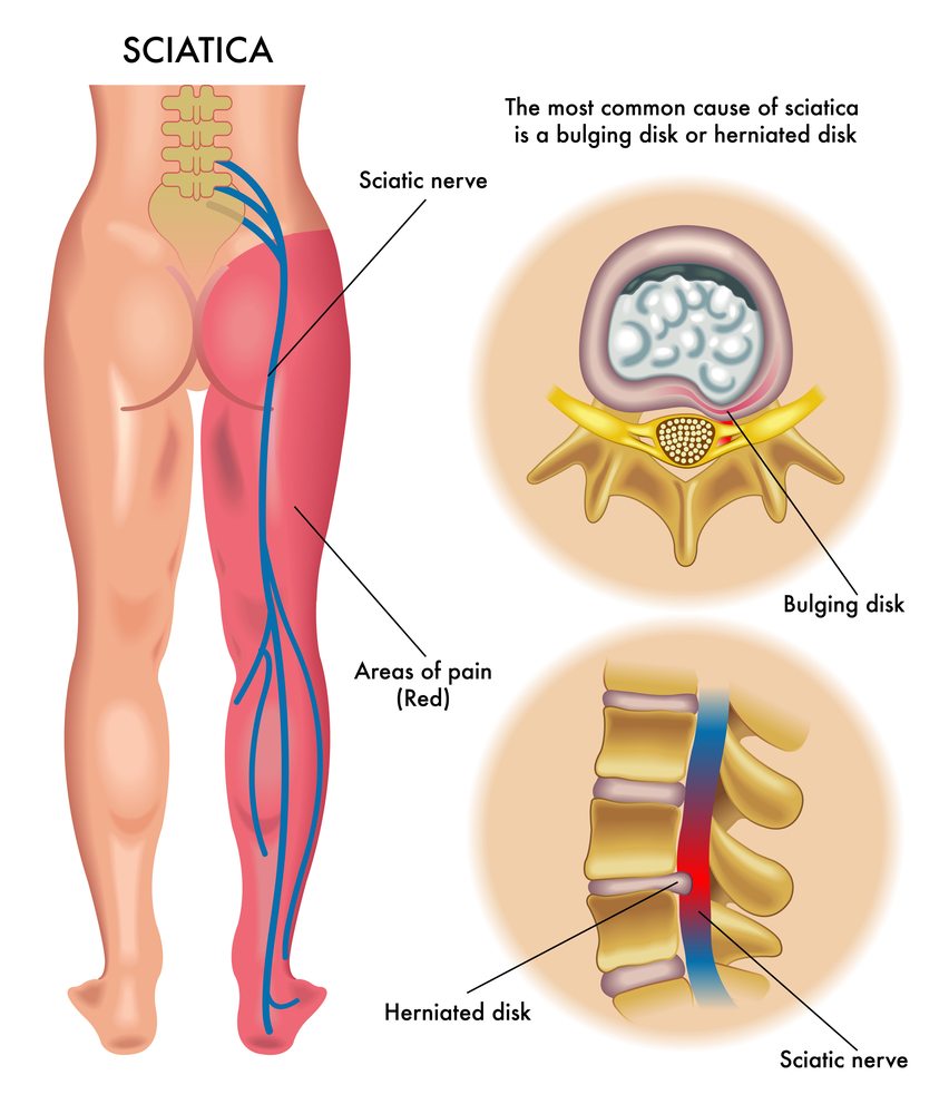
Sciatica
Sciatic nerve conduction in a high fat DMSO — Dove Medical Press
Introduction
The condition known as diabetes mellitus (DM) is a major health problem that is common throughout the modern world.1 It has been established by epidemiological research that people with diabetes are more susceptible to peripheral and central nervous system changes.2 A majority of people with diabetes suffers from the pain that is caused by diabetic neuropathy (DN).3-5 The clinical manifestations of DN are a weak peripheral nervous system, which is characterized by a reduced nerve conduction speed (NCV) leading to the appearance of signs and symptoms, such as sensation loss or painfulness in feet.6,7 Neuropathy pain can result in depression and anxiety and can cause a decline in quality of life.8
There is a gap in the treatment of DN and, therefore, its prevention is an essential aspect of diabetic care.9 The approved medications for pain relief in DN comprise Pregabalin Duloxetine as well as gabapentin, which produce an analgesic effect through activation of the a2d channel.10 However, these drugs have been associated with reported adverse reactions like dizziness, peripheral edema and somnolence.11,12 Contrary to modern medicine, botanical formulations have less adverse effects.13 The natural plant extracts are being studied for their positive effects on preclinical models for diabetes.14 Shah et al have shown the antihyperglycemic effects of Rhinacanthus Nasutus leaf extract by through in vitro and in the vivo approaches.15,16 A rhinacanthins rich R. nasutus extract was reported to effectively ameliorate the complications of diabetic nephropathy by mitigating the oxidative stress and inflammation in streptozotocin (STZ)-nicotinamide-induced model rats.17 In another study, rosemary extract was demonstrated to exert antihyperalgesic and neuroprotective effects in STZ-induced diabetic model rats.18
Black pepper from Piper nigrum L., is one of the major spices valued across the globe for its characteristic flavor.19 Black pepper fruits are a rich source of pharmacologically active terpenes, alkaloids, flavones and lignans.20 The black pepper oil contains b-caryophyllene, a CB2 (cannabinoid type 2 receptor) agonist with potential pharmacological activities reported including neuroprotection, anti-inflammation and antinociception.21-24 BCP is reported to have beneficial effects in ameliorating dyslipidemia and hyperglycemia.25 In a recent study from Geddo et al,26 the black pepper extract containing higher content of BCP was reported to have antiadipogenic activity. The extract may significantly decrease the amount of lipids that accumulate in 3T3 cells. In this study, we used an extract of the black pepper plant (Viphyllin TM) that has a standardized BCP content. We have previously studied the cognitive and neuroprotective advantages from Viphyllin. 27 Additionally, using preclinical models of pain we have confirmed the antinociceptive properties from Viphyllin. 28 To our best knowledge, the current study is the first that examines the antihyperglycemic properties of the black pepper seed extract which is standardized to the composition of BCP and its effect on nerve conduction speed in diabetic rodents.
Materials and Methods
Plant Extract
Viphyllin ViphyllinTM is a unique herbal extract obtained from Vidya Herbs Pvt. Ltd., Bangalore, India. The extract was extracted from black pepper seeds that are of Indian origin using supercritical fluid extraction. It was then standardized to the amount of b-caryophyllene that is not less than 30 percent w/w (GCMS test). 27
Chemicals and Reagents
The high-fat diet of rodents (D12492) was purchased through Research Diets, Inc. USA. Streptozotocin (STZ) (Cat No. 14653) was obtained at Sisco Research Laboratories. Ltd. (SRL), India. Glucose meter as well as glucose strips were obtained from Accu-Chek ExtraCare Roche Diabetes Care India. Ltd. Kits for biochemical analysis to measure the total amount of cholesterol and triglycerides, and LDL-cholesterol test kits were acquired at Randox Laboratories India Pvt Ltd. Creatinine, uric acids and urine test kits were bought through Robonik India Pvt Ltd. Superoxide dismutase (SOD E-BC-K020-M) catalase (CAT E-BC K031-M) and glutathione oxidase (GPx E-BC K096-M) kits were ordered by Elabscience Biotechnology Inc, USA.
Animals
Five male Wistar albino rats that were healthy and healthy with a weight of between 180 and 200 g were given to Biogen Laboratory Pvt Ltd animal house in Bangalore, India. The animals were housed in cages made of polypropylene with 12/12 h of light/dark cycles and had access to drinking water as well as a standard lab diets at 23+2 degC in rooms with a relative humidity 45-55 55%. Prior to the experiments the animals were acclimatized to the laboratory environment during seven days. The experimental protocol was reviewed and approved by the Institutional Animal Ethics Committee (IAEC) of Vidya Herbs Pvt Ltd, Bangalore, India (VHPL/PCL/IAEC/15/2020). The animal experiment protocol was executed in compliance with the Committee for the Purpose of Control and Supervision of Experiments on Animals (CPCSEA), Govt of India.
The Induction by Diabetes and treatment Schedule
The rats were split into two diets that were fed normal chow, or with HFD (D12492, Research Diets Inc. USA) for 4 weeks. Then, the overnight fasted HFD rats received only one dosage of STZ (60 mg/kg i.p., dissolving within 100mM citrate buffer pH 4.5) and the control rats were fed the drug in a single dose. Blood glucose levels in the fasting state (FBG) was determined using an Accu-chek instant-glucometer after one week of the induction period by collecting a few drops blood from the retro-orbital area under anaesthesia gaseous and rats with FBG over 200 mg/dL were classified as diabetic and were used for the study. 29
The rats in the experiment were assigned into five groupings (n=6). The normal group received Chow diet and received vehicle. Modified group/diabetic controls (HFD and STZ) group rats were fed with HFD and also received vehicle. The three groups receiving treatment received 25 100, 50 and 100 mg/kg/day doses of Viphyllin and HFD for six weeks. The weight of the animals was monitored every week. Figure 1 illustrates the experimental plan and the treatment regimen.
|
Figure 1. Treatment and experimentation timetable. |
Oral Glucose Test (OGTT)
Following the completion of the treatments, OGTT was performed in rats that were fasted overnight. The animals were given one dose of 10% glucose solution (1g/kg) via oral gavage. A few blood samples were taken from the plexus of the retro-orbital region prior to (0 minutes) and following the glucose test (30 60, 60 90, 120, and 30 minutes) and the glucose levels were measured with a the glucometer.
Test of Tail Immersion
In the study, the degree of thermal hyperalgesia in rats was evaluated using a an experiment with a tail immersion. In short, a temperature-controlled heated water bath utilized to provide warm water with temperatures of 52 + 0.5 degC. The rats were restrained gently by a towel. Then, the tail’s length of 5 cm was marked to allow the exposure to hot water. The length that was marked on the tail was submerged in warm water and the time of the tail’s immersion as well as the time of onset of the flick reaction were measured by using the stopwatch. To protect the tissue from injury the duration of cut-off was set at 30 seconds. The experiment was repeated three times per animal, with a 5 minute experimental break during trials. 30
Measurement of the Velocity of Nerve Conduction
The nerve conduction velocity (NCV) was measured at the end of the 6-week treatment duration like the one described previously. In short, the animals were anesthetized by an intraperitoneal injection ketoamine/xylazine (87 mg/13 mg b.w.). Sciatic nerve on the left leg was exposed along with electrodes (AD Instruments, PowerLab, Bella Vista, NSW, Australia) to record and stimulate the nerve were inserted in its vicinity. The action potential of the distal portion of the sciatic nerve after stimulation near the proximal point using square-wave pulses (duration: 0.001ms, intensity 200mV) which were transmitted through two electrodes recording bipolar signals was documented. A distance of 5 millimeters was maintained between stimulation electrodes. Distance between recording and stimulation electrodes, in addition to the sciatic nerve’s action-potential delay, were measured for the purpose of determining the nerve conduction rate using the formula below The NCV (m/s) will equal distance(D)/potential latency(L). 31
Biochemical Analysis
At the conclusion of the experiment the blood samples were taken for the purpose of separating serum spinning the blood samples with 1500 grams in 10 minutes. Samples of serum were kept at -80 degrees Celsius until further analysis. The samples were later examined on the basis of to determine total triglyceride (TG) as well as the total cholesterol (TC) as well as cholesterol with low density (LDL-C) and urea. blood nitrogen from urea (BUN) and uric acid levels in the chemistry analyzer of a clinical laboratory (Randox RX Imola) with commercial kits.
Organ Indices
The kidneys and the liver were quickly removed out of the visceral cavity the rat after euthanasia. They were then weighted to determine the index of organs (mg/g).
The determination of enzyme-based antioxidants on Sciatic Nerve Tissue
Sciatic nerve tissue was homogenized with an aqueous buffered with phosphate (pH 7.4) then then centrifuged to 10,000g over 15 min in the refrigerated centrifuge. The supernatants were utilized for the measurement of antioxidant enzymes’ activities. The tests were carried out using commercially available kits in accordance with the instructions of the manufacturer.
Histopathology
The kidney, liver and pancreas tissues were fixed using a 10 percent formalin solution. The paraffin-embedded samples (5 inches) were stained with the eosin and hematoxylin (H&E).
Statistics Analysis
They were then statistically analysed through one-way an analysis of variance (ANOVA) then Tukey’s test employing GraphPad Prism 9.0, and the results were presented as mean+SD. p<0.05 was considered to be statistically significant.
Results
The effect of Viphyllin on the body weight and organ indexes in diabetic Rats
Figure 2A illustrates the body mass measurements over the course of a week of the rats in the experiment. The HFD-fed rats had significantly higher weight gain between weeks 0-4 as compared to regular diet-fed control rats. However, the differences weren’t significant. After STZ induction, diabetic rats displayed a significant reductions in body weight over the course of 6 weeks to the end of the study, in comparison to the controls ( p<0.001). Viphyllin levels of 25-50 and 100 mg/kg dramatically increased their body mass of rats suffering from diabetes when compared to control rats that were not treated for diabetes ( p<0.05).
|
Figure 2. Effects of Viphyllin on body weight and organ indexes in rats suffering from diabetes. ( A) The body weight was measured weekly. The data were analysed using two-way ANOVA followed by Tukey’s Test. ( B and C) Changes in the kidney and liver indexes. The data were analysed using one-way ANOVA and Tukey’s test. The values are expressed as mean +-SD (n=5). #p<0.05, ##p<0.01 and ###p<0.001 against control *p<0.05, **p<0.01 and ***p<0.001 against control for diabetes. Abbreviations: ANOVA, analysis of variance and SD, standard deviation. |
Diabetic rats had significantly greater liver (55.85 percent) and kidney indexes (90.81 percent) than the control groups ( p<0.001). However treatment with Viphyllin in different doses significantly increased the organ indices in diabetes-related rats (Figure 2B). Viphyllin doses of 25-50 and 100 mg/kg demonstrated an increase of 20.51 percent ( p<0.001), 17.63% ( p<0.01) and 12.83 percent ( p<0.05) in the liver indices , respectively when compared with the diabetic untreated rats. The Viphyllin-treated rats further decreased the kidney indices by 21.20 percent ( p<0.01), 20.14% ( p<0.01) and 25.36 percent ( p<0.001) in comparison to diabetic rats that were not treated (Figure 2C).
The effect of Viphyllin on oral Glucose Tolerance and fasting blood Glucose (FBG) within diabetic Rats
Blood glucose levels (BGL) was substantially more elevated in the control diabetic group at 0 , and then increased significantly upon the administration of glucose (1 mg/kg, p.o.) until 120 min as compared to the control rats ( p<0.001). Viphyllin treatment at various doses reduced BGL in diabetic rodents for 120 minutes. Viphyllin dosages of 50 ( p<0.05) and 100 mg/kg ( p<0.001) significantly decreased BGL within 60 min after glucose injection in rats with diabetes as compared to the diabetic control sample (Figure 3A). AUC analysis showed the following: Viphyllin treatments at 25 mg/kg, and 100 mg/kg revealed 5.42 percent, 11.77% ( p<0.05) and 12.11 percent ( p<0.05) reduction in BGL levels, respectively when compared with diabetes control animals (Figure 3B). As one would expect, there was an increase in FBG in diabetic rats at the end of the study when compared with the control ( p<0.001). Viphyllin 100 mg/kg significantly reduced FBG when compared to diabetic rats that were not treated ( p<0.05) (Figure 3C).
|
3. Effects of Viphyllin on the tolerance to glucose for diabetic rodents. ( A) Blood glucose levels determined at the time of zero and following the oral administration of (1 grams/kg mass) glucose. ( B) Area under the curve (AUC glucose) between 0 and 120 min following the administration of glucose by mouth. ( C) The blood glucose level at the end of the study. The data were analyzed using one-way ANOVA and Tukey’s test. Values are expressed as mean+SD (n=5). ##p<0.05 and ###p<0.001 against control *p<0.05 and ***p<0.001 in comparison to diabetic control. Abbreviations: ANOVA, analysis of variance SD, standard deviation. |
The effect and effect of Viphyllin Effect of Viphyllin Thermal Hyperalgesia in the Diabetic Rats Model
In Figure 4 there was a noticeable decrease in the tail flick latency duration observed in diabetic rats as compared to the controls ( p<0.001) that indicates the presence of thermal hyperalgesia. However administration of Viphyllin at doses of 25, 50 or 100 mg/kg in dose dependent manner, increased the duration of latency in comparison to the diabetic groups ( p<0.05).
|
Figure 4. Impact of Viphyllin on the latency of tail flicks of diabetic rats. The data were analysed using one-way ANOVA and Tukey’s test. The values are expressed as mean+SD (n=5). #* * p<0.001 against control * p<0.05 in comparison to diabetic control. Abbreviations: ANOVA, analysis of variance and SD, standard deviation. |
The effect on Viphyllin Effect of Viphyllin Nerve Conductance Velocity and the Antioxidant Enzyme’s Activity of Sciatic Nerve Tissue
The diabetic rats displayed significant ( p<0.001) lower the NCV value (25.80+-1.88 m/s) when compared with animal control group (55. +-6.46 m/s). A six-week treatment with Viphyllin produced 12.95 percent, 30.70% and 71.01 percentage increase in the NCV at 25 50 ( p<0.05) and 100 mg/kg ( p<0.001) dosages, respectively, when compared to the control for diabetes (Figure 5A).
|
Figure 5. Influence of Viphyllin nerve conduction speed (NCV) ( A) and antioxidant condition ( B– D) in sciatic nerve tissue. The data were examined using one-way ANOVA and Tukey’s test. The values are expressed as mean+SD (n=5). #p<0.05 and ###p<0.001 against control *p<0.05, **p<0.01 and ***p<0.001 against diabetic control. Abbreviations: ANOVA, analysis of variance SD, Standard deviation. |
The diabetic model rats displayed significant decreases in SOD ( p<0.05) as well as GPx, CAT and ( p<0.001) levels of enzymes within the sciatic nerve tissue when compared with rats that were not diabetic (Figure 5B-D). Viphyllin dramatically improved the enzyme activity within diabetic animals. At 100 mg/kg Viphyllin demonstrated 1.37-fold ( p<0.05), 3.73-fold ( p<0.001) and 1.15-fold increase in SOD, level of GPx and CAT when compared with untreated diabetic rats.
The effect of Viphyllin administration On Serum Biochemical Markers in Diabetic Rats
As illustrated in Figure 6. evident increase of TCA ( p<0.05) and the TG ( p<0.001) and LDL-c levels of diabetic rats as compared to the controls. In contrast, Viphyllin dose-dependently reduced the TC and TG levels in diabetic rats. The TC decreased to 22.86 percent ( p<0.05) and 31.31 percent ( p<0.001) in 50 as well as 100 mg/kg Viphyllin treatment groups, compared to the diabetic control. The doses of 100 mg and 50 mg/kg dramatically reduced the TG levels (33.86 percent and 40.69 percent respectively) in comparison to the diabetic animals ( p<0.001). Viphyllin treatments also reduced levels of LDL-c in the serum. However, the results did not show any significant difference when compared to the diabetic rats who were not treated.
|
Figure 6. Effects of Viphyllin on the serum cholesterol profile of diabetic rats. The data were examined using two-way ANOVA and reported in terms of mean+SD (n=5). The values are mean+SD (n=5). #p<0.05 and ###p<0.001 against control *p<0.05 and ***p<0.001 in comparison to diabetic control. Abbreviations: ANOVA, analysis of variance SD, standard deviation. |
The diabetics who were untreated had significantly higher levels of creatinine ( p<0.001) and uric acid ( p<0.05) when compared to the control rats, which indicates kidney damage. Viphyllin treatment dramatically reduced renal health indicators among diabetic animals (Figure 7A and B). 100 mg/kg Viphyllin demonstrated 38.24 percent and 58.16 percent reduction in serum levels of uric acid and creatinine and uric acid, respectively, compared to untreated diabetic rodents ( p<0.01). Additionally, Viphyllin insignificantly lowered the BUN levels of the diabetic rodents (Figure 7C).
|
Figure 7. Effects of Viphyllin on the levels of serum kidney health indicators among diabetic rats. ( A) Creatinine, ( B) the uric acid as well as ( C) blood nitrogen from urea. The data were analysed using one-way ANOVA and Tukey’s test. The values are mean+SD (n=5). #p<0.05 and ###p<0.001 in comparison to control *p<0.05 and **p<0.01 against control for diabetes. Abbreviations: ANOVA, analysis of variance SD, standard deviation. |
The effect and Effect of Viphyllin Effect of Viphyllin Histopathological changes in Liver, Kidney, and Pancreas
The histopathological examination of livers showed normal hepatocytes in the control group , while in diabetic control group , the characteristic changes in pathology including. inflammation, inflammation foci and fat vacuolations, as well as hyperplastic biliary tissue were apparent. Viphyllin dosages of 25, 50 , and 100 mg/kg significantly improved liver’s structure for diabetic rodents (Figure 8A). The diabetic rats displayed STZ-induced changes like glomerular atrophy, inflammation, arterial fibrosis and the vacuolation of fats. However, Viphyllin treated diabetic rats were able to reverse these pathological changes and restored regular theomorphology (Figure 8B). The rats in the control group showed the normal pancreas’ structure with normal arelets from Langerhans and acinar cell whereas the diabetic control rats displayed symptoms of pathology such as swelling of Acinar cells, emptying of islets and atrophy of the islets as well as degenerative hyperplasia as well as congestion. It is interesting to note that Viphyllin treated rats had normal pancreas structure (Figure 8C).
|
Figure 8. Effects of Viphyllin on histopathology of vital organs of rats suffering from diabetes. Images representative (H&E staining) showing histological changes in the liver ( A) and kidney ( B) and the pancreas ( C). The green arrowhead indicates inflammation. green arrows signify hyperplastic biliary tissue Yellow arrowhead normal glomeruli; yellow the arrows indicate glomerular atrophy. black arrowhead normal islets in Langerhans White arrowhead in the form of atrophy of the islets of Langerhans Black arrow emptying islets and white arrows indicate congestion. (Magnification: x100; scale bar: 100 um). |
Discussion
This study was carried out to study the therapeutic effects that are attributed to Viphyllin as an experiment in a model that mimics diabetes. HFD that is a result of a only one doses of STZ has been previously documented to establish type 2 diabetes in laboratory animals. 32,33 HFD is a factor in the development of insulin resistance, while STZ causes impaired insulin secretion. 34 In this study the female Wistar rats received HFD for four weeks, and then received intraperitoneal injections of STZ in the amount of 60 mg/kg. The rats that were fed HFD had an insignificant increase in body weight over the initial 4 weeks of the study. However, the following STZ treatment led to weight loss as well as increased BGL when compared the normal rodents. However, the 6-week oral treatment with Viphyllin significantly improved body weight as well as FBG, in rats suffering from diabetes. Viphyllin treatment reduced STZ-induced rise in kidney and liver indexes.
It is a test to determine diminished in the ability to detect insulin. 35 In the current study, diabetic rats showed significant higher levels of BGL during 120 minutes of glucose load when than control rats, indicating the resistance to insulin. In addition, the AUC of glucose however was diminished significant for Viphyllin treated rats in comparison to control rats that were not treated for diabetes. These findings clearly show the antihyperglycemic properties of Viphyllin.
DM related nerve conduction problems and neuropathic pain can cause changes in the sensory responses. 36 The shift in the sensation and neuropathic pain causes hyperalgesia as well as allodynia. 37 Several studies have demonstrated the power of plant extracts in regulating the threshold for pain of the models of neuropathic pain. 38,39 Behavioral examination of an altered responses to external stimuli is a clear indicator of neuropathological changes in people suffering from diabetes. In the current study, diabetic rats displayed an increase in the latency of thermal stimuli (tail flash test) that indicated hyperalgesia. In contrast, Viphyllin treatment notably increased the duration of the tail flick in diabetic rats to an impressive degree. The nerve conduction problems that is associated with neuropathy was evident in diabetic rats. The NCV was significantly decreased in diabetic rats that were not treated when compared to controls, while Viphyllin treatment rats demonstrated improvements in sciatic nerve conduction. The results of this study is in line with earlier studies. In the past, Laddha et al demonstrated the connection with the antinociceptive action from Bauhinia variegata extracts in the context of increased NCV in rats suffering from diabetes. 40 In another study that included an ayurvedic remedy, Triphala churna significantly increased the duration of the nociceptive reaction in diabetic models. Additionally, the researchers showed that Triphala significantly increased the level of NCV in rats. 41
Biochemically, mammalian nerve tissues contain phospholipids as well as a high number of mitochondria that generate sufficient free radicals to trigger an oxidative injury. 42 Previous research has demonstrated that a weak antioxidant defense in nerve tissues can cause neuropathic pain on animal models. 43 In keeping with these findings the diabetic rats demonstrated an inactive antioxidant enzymes within the sciatic nerve in comparison to normal rats. Viphyllin greatly improved the antioxidant defense of sciatic nerve tissue of rats. The Viphyllin-mediated enhancement of NCV and the activity of antioxidants on the sciatic nerve may be due to its ability to modulate peripheral sensory functions.
Diabetic complications eventually cause renal problems along with hyperlipidemia. 44,45 In the current investigation, Viphyllin treatment improved the levels of serum lipids and the renal parameters of diabetic rats. The histopathological changes that occurred in the vital organs like pancreas, kidney, and liver in diabetic rodents were reversed an excellent extent through the treatment with Viphyllin. These results further confirm the antiidiabetic effects of Viphyllin.
A substantial amount of research findings confirm the therapeutic value for BCP, the well-studied active component of Viphyllin. 46 Previous studies, Suijin et al have found that treating pancreatic b cells by BCP increased insulin-stimulated glucose release. 47 BCP as the main component in various plant extracts has been shown to be hypoglycemic for diabetic rodents. 48 Recently Aguilar-Avila and colleagues examined the effects of BCP on the neuropathic pain of diabetic rats. BCP can significantly reduce BGL while also increasing insulin levels in diabetics treated with STZ. 49 These results strongly suggest the role of BCP in the Viphyllin-mediated therapeutic effect in the current study.
Conclusion
The oral administration of Viphyllin significantly improved sciatic nerve conduction and sensorimotor responses in diabetics induced by HFD/STZ. Furthermore the extract showed antihyperglycemic properties in diabetic rats. The current research results suggest that there are protection effects of Viphyllin in neuropathy caused by diabetes.
Acknowledgment
The authors are grateful to their doctor Dr. Muralidhar for technical assistance during the histopathological examination of the tissues.
Disclosure
All authors are employees of Vidya Herbs Pvt. Ltd. They therefore declare any potential conflicts of interest The authors do not report any conflict of interest relating to this research.
References
1. International Diabetes Federation.
IDF Diabetes Atlas
.
8th
ed. Brussels, Belgium: International Diabetes Federation 2017.
2. Vinik AI, Nevoret M-L, Casellini C, et al. Diabetic neuropathy. Endocrinol Metab Clin North Am. 2013;42:747-787. doi:10.1016/j.ecl.2013.06.001
3. Varkonyi T, Korei A, Putz Z, et al. The latest advances in the treatment of neuropathy caused by diabetes. Minerva Med. 2017;108:419-437. doi:10.23736/S0026-4806.17.05257-0
4. Callaghan BC, Price RS, Chen KS, et al. The significance of subtypes with rare characteristics in the the diagnosis of and management for peripheral neuropathy. A review. JAMA Neurol. 2015;72:1510-1518. doi:10.1001/jamaneurol.2015.2347
5. Abbott CA, Malik RA, van Ross ER, et al. The prevalence and characteristics of painful diabetic neuropathy a large population of diabetics living in communities within the U.K. Diabetes Care. 2011;34:2220-2224. doi:10.2337/dc11-1108
6. Tesfaye S, Boulton AJ, Dyck PJ, et al. Diabetic neuropathies: updates on definitions as well as diagnostic criteria, estimation of severity, as well as treatments Corrections published within Diabetes Care. Diabetes Care. 2010;33:2285-2293. doi:10.2337/dc10-1303
7. von Hehn CA, Baron R, Woolf CJ. The neuropathic pain phenotype is deconstructed to discover neural mechanisms. Neuron. 2012;73:638-652. doi:10.1016/j.neuron.2012.02.008
8. Kioskli K, Scott W, Winkley K, et al. Psychosocial aspects in painful diabetic neuropathy: A systematic review of treatment trials as well as surveys. Chronic Pain Med. 2019;20:1756-1773. doi:10.1093/pm/pnz071
9. Pop-Busui R, Boulton AJ, Feldman EL, et al. Diabetic neuropathy: A position statement issued by the American diabetes association. Diabetes Care. 2017;40:136-154. doi:10.2337/dc16-2042
10. Javed S Alam U Malik RA. Mirogabalin and new therapies in the treatment of diabetic neuropathy. J Pain Res. 2018;11:1559-1566. doi:10.2147/JPR.S145999
11. Freeman R, Durso-Decruz E, Emir B. Efficacy as well as safety and tolerability of pregabalin therapy for peripheral neuropathy in patients with painful diabetes Seven randomized controlled trials over various dosages. Diabetes Care. 2008;31:1448-1454. doi:10.2337/dc07-2105
12. Wiffen PJ, Derry S, Bell RF, et al. Gabapentin for chronic nerve pain in adults. Cochrane Database Syst Rev. 2017;6:Cd007938. doi:10.1002/14651858.CD007938.pub4
13. Tiwari R, Siddiqui MH, Mahmood T, et al. Herbal remedies: a boon to diabetic neuropathy. J Diet Suppl. 2019;16:470-490. doi:10.1080/19390211.2018.1441203
14. Lee J, Noh S, Lim S, et al. Extracts from plants for type 2 diabetes The journey from traditional treatment to current drug discovery. Antioxidants. 2021;10(1):81. doi:10.3390/antiox10010081
15. Shah MA, Reanmongkol W, Radenahmad N, et al. Anti-hyperglycemic and anti-hyperlipidemic effects of rhinacanthins-rich extract from Rhinacanthus nasutus leaves in nicotinamide-streptozotocin induced diabetic rats. Biomed Pharmacother. 2019;113:108702. doi:10.1016/j.biopha.2019.108702
16. Shah MA, Khalil R, Ul-Haq Z, et al. a-Glucosidase inhibitory effects of rhinacanth extracts from Rhinacanthus Nasutus leaf and synergistic effects when combined with the acarbose. J Funct Foods. 2017;36:325-331. doi:10.1016/j.jff.2017.07.021
17. Zhao LL, Makinde EA, Shah MA, Olatunji OJ, Panichayupakaranant P. Rhinacanthins-rich extract and rhinacanthin C ameliorate oxidative stress and inflammation in streptozotocin-nicotinamide-induced diabetic nephropathy. J Food Biochem. 2019;43(4):e12812. doi:10.1111/jfbc.12812
18. Rasoulian B, Hajializadeh Z, Esmaeili-Mahani S, et al. Antinociceptive and neuroprotective effects on rosemary ( Rosmarinus officinalis L.) extract in diabetic rats suffering from painful neuropathy. J Physiol Sci. 2019;69:57-64. doi:10.1007/s12576-018-0620-x
19. Salehi B, Zakaria ZA, Gyawali R, et al. Piper species: A complete review of their phytochemistry, biological functions and potential applications. Molecules. 2019;24:1364. doi:10.3390/molecules24071364
20. Musenga A, Mandrioli R, Ferranti A, et al. Analysis of aromatic and terpenic constituents of pepper extracts by capillary electrochromatography. J Sep Sci. 2007;30:612-619. doi:10.1002/jssc.200600456
21. Viveros-Paredes JM, Gonzalez-Castaneda RE, Gertsch J, et al. Beta-caryophyllene’s neuroprotective properties against dopaminergic neuron injuries in a model mouse of Parkinson’s disease caused by MPTP. Pharmaceuticals. 2017;10:60. doi:10.3390/ph10030060
22. Guo K, Mou X, Huang J, et al. Trans-caryophyllene reduces neuroinflammatory reactions induced by hypoxia by suppressing the NF-kappa B stimulation in microglia. J Mol Neurosci. 2014;54:41-48. doi:10.1007/s12031-014-0243-5
23. Ojha S, Javed H, Azimullah S, et al. beta-caryophyllene is a phytocannabinoid decreases neuroinflammation, oxidative stress, the activation of glia, and helps to salvage dopaminergic cells in a rat model for Parkinson disease. Mol Cell Biochem. 2016;418:59-70. doi:10.1007/s11010-016-2733-y
24. Klauke AL, Racz I, Pradier B, et al. Cannabinoid CB2 receptor-selective phytocannabinoid Beta-caryophyl analgesic effects on mice models of neuropathic and inflammatory pain. Eur Neuropsychopharmacol. 2014;24:608-620. doi:10.1016/j.euroneuro.2013.10.008
25. Kumawat S, Kaur G. Potential therapeutic value of the cannabinoid receptor 2 treating diabetes and complications. Eur J Pharmacol. 2019;862:172628. doi:10.1016/j.ejphar.2019.172628
26. Geddo F, Scandiffio R, Antoniotti S, et al. PipeNig(r)-FL, a fluid extract of black pepper (Piper nigrum L.) with a high standardized content of Trans-b-caryophyllene, reduces lipid accumulation in 3T3-L1 preadipocytes and improves glucose uptake in C2C12 myotubes. Nutrients. 2019;11:2788. doi:10.3390/nu11112788
27. Sudeep HV, Venkatakrishna K, Gouthamchandra K, et al. A standardized extract of black pepper seeds that contains b-caryophyllene enhances cognitive function in scopolamine-induced amnesia models by regulating neurotrophic factor that is derived from the brain as well as MAPK proteins. J Food Biochem. 2021;45:e13994. doi:10.1111/jfbc.13994
28. Venkatakrishna K, Sundeep K, Sudeep HV, et al. Viphyllin, a standardized black pepper seed extract exerts antinociceptive effects in murine pain models via activation of cannabinoid receptor CB2, peroxisome proliferator-activated receptor-alpha and TRPV1 ion channels. J Pain Res. 2022;15:355-366. doi:10.2147/JPR.S351513
29. Zhang M, Lv XY, Li J, et al. The characterisation of a the high-fat diet as well as streptozotocin in multiple doses that cause type 2 diabetes in a rat model. Experiment in Research in Diabetes. 2008;2008:704045. doi:10.1155/2008/704045
30. Leng J, Li X, Tian H, et al. Diosgenin’s neuroprotective properties in a mouse model of diabetes peripheral neuropathy is mediated by an Nrf2/HO-1 signaling pathway. BMC Complement Med Ther. 2020;20:126. doi:10.1186/s12906-020-02930-7
31. Wang X, Huan Y, Li C, et al. Diphenyl diselenide alleviates diabetic peripheral neuropathy in rats with streptozotocin-induced diabetes by modulating oxidative stress. Biochem Pharmacol. 2020;182:114221. doi:10.1016/j.bcp.2020.114221
32. Atanasovska E, Tasic V, Slaninka-miceska M, et al. Six-week observation of the metabolic effects caused by a high-fat diet as well as streptozotocin, a model rodent for the disease type 2. Contributions Sec Med Sci. 2014;35:169-179.
33. Veerapur VP, Prabhakar KR, Kandadi MR, et al. Antidiabetic effect of Dodonaea viscosa aerial parts in high fat diet and low dose streptozotocin-induced type 2 diabetic rats: a mechanistic approach. Pharm Biol. 2010;48:1137-1148. doi:10.3109/13880200903527736
34. Srinivasan K, Viswanad B, Asrat L, et al. Combination of high-fat diet-fed and low-dose streptozotocin-treated rat: a model for type 2 diabetes and pharmacological screening. Pharmacol Res. 2005;52:313-320. doi:10.1016/j.phrs.2005.05.004
35. Shaham O, Wei R, Wang TJ, et al. Metabolic profiling of reaction of the human body to glucose-related challenge shows distinct areas in the sensitivity to insulin. Mol Syst Biol. 2008;4:214. doi:10.1038/msb.2008.50
36. Tesfaye S, Boulton AJ and Dickenson A.H. Mechanisms and treatment of diabetic painful distal polyneuropathy symmetrical. Diabetes Care. 2013;36:2456-2465. doi:10.2337/dc12-1964
37. Didangelos T, Doupis J, Veves A. Neuropathy with pain Clinical aspects. Handbook Clin Neurol. 2014;126:53-61.
38. Wang P, Wen C, Olatunji OJ. Anti-inflammatory and antinociceptive properties of Boesenbergia Rotunda Polyphenol in peripheral diabetic neuropathy rats. J Pain Res. 2022;15:779-788. doi:10.2147/JPR.S359766
39. Nawaz NUA, Saeed M, Rauf K, et al. Effectiveness of the antinociceptive properties in Tithonia tubaeformis in a vincristine-based model of chemotherapy-induced pain neuralgia in mouse. Biomed Pharmacother. 2018;103:1043-1051. doi:10.1016/j.biopha.2018.04.115
40. Laddha AP, Garud MS, Kulkarni YA. Neuroprotective properties from Bauhinia variegata Linn. Leaf extracts in streptozotocin-induced the development of diabetes Sprague Dawley rats. J Diabetes Metab Disord. 2021;20(2):1639-1645. doi:10.1007/s40200-021-00915-y
41. Suryavanshi SV, Barve K, Addepalli V, et al. Triphala Churna, a traditional formula in Ayurveda that reduces neuropathy caused by diabetes in rodents. Front Pharmacol. 2021;12:662000. doi:10.3389/fphar.2021.662000
42. Areti A, Yerra VG, Naidu V, et al. The effects of oxidative stress on nerve damage The role of chemotherapy in peripheral neuropathy. Redox Biol. 2014;2:289-295. doi:10.1016/j.redox.2014.01.006
43. Kawaguchi M, Satoh Y, Otsubo Y, et al. Molecular hydrogen can reduce neuropathopathic pain in mice. The journal PLoS One. 2014;9:e100352. doi:10.1371/journal.pone.0100352
44. Yokota K, Fukuda M, Matsui Y, et al. Variation in blood pressure from visit to visit and decline in renal function in patients suffering from chronic kidney disease that is diabetic. J Clin Hypertens. 2014;16:362-366. doi:10.1111/jch.12293
45. Bhowmik B, Siddiquee T, Mujumder A, et al. Serum cholesterol profile and its connection with prediabetes and diabetes in the Rural Bangladeshi population. int J Environ Res Public Health. 2018;15(9):1944. doi:10.3390/ijerph15091944
46. Hashiesh HM, Meeran MFN, Sharma C, Sadek B, Kaabi JA, Ojha SK. The therapeutic potential of b-caryophyllene is a cannabinoid found in food in diabetes and its complications. Nutrients. 2020;12:2963. doi:10.3390/nu12102963
47. Suijun W, Zhen Y, Ying G, et al. Trans-caryophyllene’s role in the modulation the secretion of insulin. Biochem Biophys Res Commun. 2014;444:451-454. doi:10.1016/j.bbrc.2013.11.136
48. Uddin N, Hasan MR, Hossain MM, et al. In in vitro inhibition of a-amylase and in vivo hypoglycemic impact of methanol extracts of Citrus macroptera Montr. fruit. Asian Pac J Trop Biomed. 2014;4:473-479. doi:10.12980/APJTB.4.2014C1173
49. Aguilar-Avila D, Flores Soto M, Tapia-Vazquez C, et al. B-caryophyllene, a naturally occurring sesquiterpene that reduces depression-like and neuropathological pain in diabetic mice that have been subjected to experimental testing. J Med Food. 2019;22:460-468. doi:10.1089/jmf.2018.0157


Understanding Leg Pain: Causes, Symptoms, and How Chiropractic Care Can Help

Are Core Strengthening Exercises Effective for Lower Back Pain Relief?

Nerve Root Compression: Understanding, Symptoms, and Effective Treatments

Understanding and Treating a Herniated Disc: Your Guide to Relief and Recovery

Lower Back Pain: What’s Really Going On and How You Can Feel Better

Understanding Sciatica and Radiculopathy: Causes, Symptoms, and Management

How Often Should You Exercise to Maintain Lower Back Health?

What Are the Top 10 Exercises for Sciatica Pain Relief?

How Does Proper Hydration Reduce Muscle Pain Naturally?

Are Mesh Back Chairs Better for Sciatica Pain Relief?

How Does Stretching Help in Easing Sciatica Discomfort?

What Are the Top 10 Herbal Remedies for Natural Muscle Pain Relief?

How Does Regular Exercise Help Alleviate Lower Back Pain?

How Does Aging Contribute to Chronic Lower Back Pain?
¿Desaparecerá el entumecimiento de los pies con ciática?

How Does Heat Therapy Provide Natural Relief From Muscle Pain?

How Effective Are Herbal Supplements for Alleviating Sciatica Pain?
The Pain Tape Game: Does KT Tape Help With Sciatica?

Will Foot Numbness with Sciatica Go Away?

What Causes Sciatica Nerve Pain
How To Get Sciatica To Stop Hurting
How To Get Rid Of Sciatica Pain During Pregnancy
How To Drive With Sciatica

Will Sciatica Show Up On MRI
How To Fix Sciatica Pain Fast
How To Help Sciatica Pain Naturally
How To Help Sciatica Pain At Home
How To Know Its Sciatica Pain